Mucosal disease, BVD
AETIOLOGY:
Pestivirus of the Flaviviridae family. RNA virus, with ability to recombine. Different serotypes. There are two byotipes of the virus, the cytopathic and the non-cytopathic form. They are only differentiable in cellular cultures by the lysis caused. Other diseases caused by pestivirus are Border disease of small ruminants and swine fever.
TRANSMISSION:
- Direct: (nasal-pharyngeal secretions, urine), aerosols and by venereal route. Faeces are a weak source of infection. The PII calf persistently (persistently infected immunotolerant) occurs when the fetus is infected before the 120th day of gestation; this is the time in which its immune system is mature and functional. The presence of PII animals in a group is key to the appearance of acute outbreaks of BVD, as it is highly infective throughout its lifetime.
CLINICAL SIGNS:
It nearly always presents slight or unapparent clinical symptoms, except in two situations: when a gestating cow is infected or when there is a co-infection with another virus, for example, those of tropism of the respiratory tract (BRSV, IBR, PI-3, Adenovirus, etc.).
- Cow in heat: breeding and infertility (of 6 to 8 weeks). Early resorptions.
- Pregnant cow: infection in the first four months of gestation may lead to still births, mummification and early foetal death. - If the fetus becomes infected and is not aborted, it will be born a lifetime carrier of BVD (PII). Infection after 120 days may harm the fetus (cerebral Hypoplasia) but it does not become immunotolerant.
- Concomitant infections: BVD induces a marked immunosuppression that exacerbates concomitant infections and, more commonly, those that are respiratory in nature.
- Mucosal disease: secondary infections of a PII animal with the cytopathic type of BVD. Diarrhoea, anorexia; buccal and nasal ulcers and ulcers on the tongue. Limping, prostration and inevitable death in 5-10 days.
LESIONS:
Oral ulcers, ulcers of the abomasum, small intestine and colon.
DIAGNOSIS:
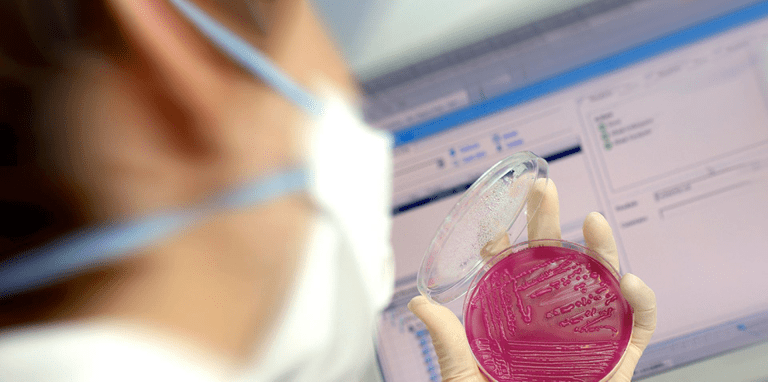
Isolation and identification - Serology: ELISA, IHA, SNT and PCR.
TREATMENT, PREVENTION AND CONTROL:
Elimination of PII animals from the group. Vaccinal prophylaxis of all animals. Live vaccines (contraindicated in gestating animals) and inactivated vaccine.