AETIOLOGY:
Erysipelothrix rhusiopathiae, gran-positive bacterium
TRANSMISSION:
30-50% of apparently healthy pigs carry the bacterium in their tonsils and other lymphoid tissues. The organism is shed in multiple excretions and secretions for extended periods of time. The transmission occurs via oronasal or skin wounds. The bacterium can remain infectious in the environment for long periods of time.
CLINICAL SIGNS:
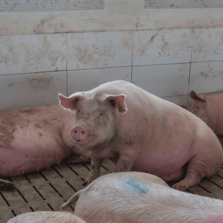
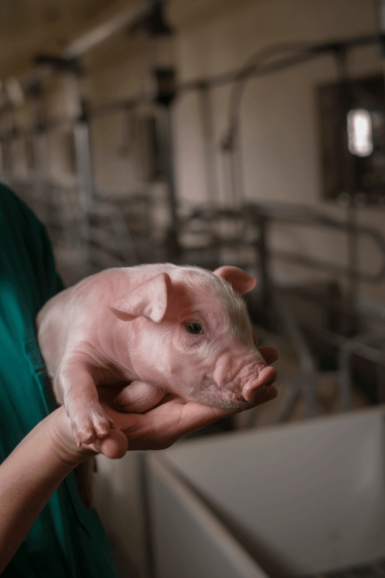
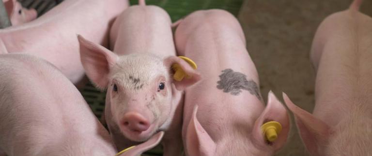
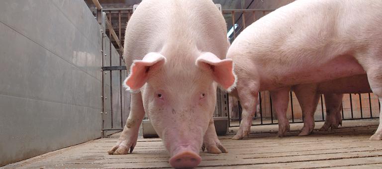
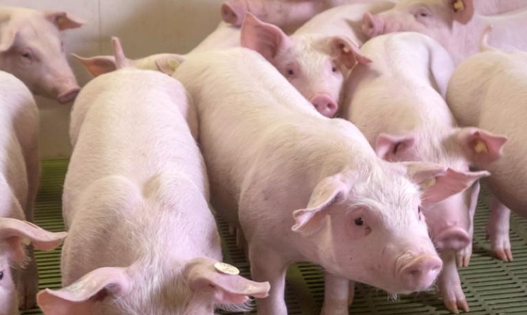
There are 3 clinical forms of swine erysipelas: acute, subacute and chronic, with a huge variety of clinical signs.. It can affects pigs commonly of 10-12 weeks onwards, sows and boars. The acute form is septicemic disease that manifests as sudden onset o acute death, abortions, depression, lethargy, pyrexia, lying down, reluctant to move and the characteristic pink, red or purple raised firm rhomboid or “diamond skin” lesions. The subacute is less severe than the acute. Animals do not appear as sick, temperature is not as high or persistent, skin lesions may be few in number of absent, there may be infertility, litters with mummies or small size, vulvar discharges, going in some cases unnoticed. The chronic form, follows the acute, subacute or subclinical in a proportion of surviving animals. The most significant sign is chronic arthritis that may appear around 3 weeks after the initial outbreak. Derived of the lameness, the affected animals may have associated reduction in feed intake, respiratory distress, lethargy, and sudden death as consequence of vegetative valvular endocarditis.
LESIONS:
The pathognomonic gross lesions consist of multifocal pink to purple diamond-shaped slightly raised skin lesions. There are other typical lesions of septicemia like congested lymph nodes, enlarged spleen, edematous and congested lungs. Regarding the arthritis, the synovium and periarticular tissues are typically distended by serofibrinous exudates. In hyperacute processes, some animals can be found dead without any gross lesions. Vegetative valvular endocarditis can be observed.
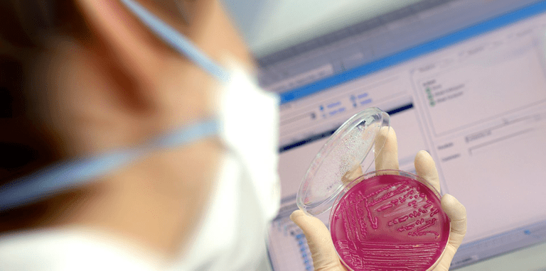
DIAGNOSIS:
Clinical: when the skin diamond shaped lesions appear. Due to the broad range of clinical symptoms, the clinical diagnosis it is not easy.
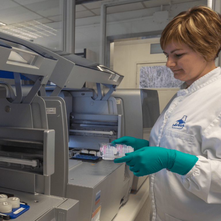
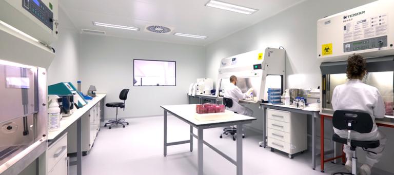
Laboratory: isolation and culture from tissues with lesions provides a definitive diagnosis. In case of chronic processes, prior selective enrichment methods are recommended. Also exist PCR methods for the rapid detection. The serological assays most common technique is the ELISA that allows to detect antibodies
TREATMENT, PREVENTION AND CONTROL:
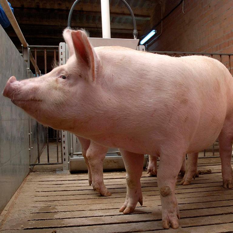
In acute cases, antimicrobial therapy it is recommended my injectable penicillin. The prevention and control must be accomplished by immunization programs. Most vaccines are inactivated bacterins, based on serotypes 1 or 2. The duration of immunity varies between 6 to 12 months. In fattening animals when necessary are injected from 10 weeks of age onwards. Breeding animals are commonly revaccinated every productive cycle to prevent reproductive disorders. The vaccines commonly contain Porcine Parvovirus and Leptospira spp. antigens, depending on the farming and epidemiological conditions.