Article written by:
Oriol Boix Mas (Corporate Product Manager at HIPRA).
Igancio Bernal Orozco (Corporate Brand Manager at HIPRA).
5b. HEALTH: How to get a successful diagnosis?
Posts
Porcs
In cases where we are faced with a gastrointestinal disorder of infectious origin, owing to their multifactorial aetiology it is of vital importance to take action in a meticulous and orderly manner.
To do this, we as veterinarians have a series of tools and procedures available to us, which allow us in the first instance to make a differential diagnosis and then determine the source of the problem.
This includes taking a correct history, performing an appropriate necropsy and taking specific samples that will allow us to request the appropriate tests from the laboratory.

Material needed for a good gastrointestinal necropsy
On a general level, the most obvious clinical manifestation will be the presence of diarrhoea, in the majority of cases accompanied by a change in the colour of the faeces.
Certainly, and especially in cases of viral infection, the presence of vomiting, mucus, blood or undigested feed are symptoms that can provide more pointers towards a presumptive diagnosis.
1. The importance of proper sampling
The success of diagnosis depends on the selection of the samples, as these must be representative and must be in a good state of preservation.
How many animals?:
- 4-5 animals from different litters. To obtain a good representation of the farm.
Which animals?:
- Animals in the initial phases of diarrhoea: Animals, which have had diarrhoea for longer, may be more difficult to diagnose because of the presence of secondary agents.
- Untreated animals: Antibiotic treatment can prevent the isolation of bacteria such as E. coli or Clostridium.
- Sacrificed animals: Avoid samples from animals that have been dead for several hours as autolysis of the intestine sets in rapidly.
Which samples?:
- Digestive tract: Part of the small intestine and of the large intestine with the ends tied. These will allow a complete analysis, including the histopathological study.
- Swabs and faeces: Whenever a digestive tract sample cannot be sent, obtain the samples directly from the rectum: insert the swab, gently rubbing it against the walls and rotating it.

Rectal sampling swab
How to collect and dispatch the samples?:
- Send the refrigerated samples to the laboratory as soon as possible (within 24 hours). Send fresh samples for microbiological examination and PCR; and in formalin for the histopathological study.
- As regards the equipment required for sampling, we need latex gloves, vials and bags with a hermetic seal and swabs with a transport medium such as AMIES or STUART in order to avoid the desiccation of the sample and the loss of bacterial viability.
- In addition, for international dispatch for sampling for PCR, FTA cards can also be used.
- Finally, a bottle of formaldehyde 10% will be needed to fix samples intended for histopathological study and white cork boxes with ice gel blocks.
2. Autolysis, the enemy of sampling and necropsy
Autolysis is the process by which the cells of the animal, once it is dead, release enzymes known as autolysins that start a process of degradation and cellular self-destruction.
When taking samples, we must bear in mind that the digestive system is characterized by its high concentration of enzymes, hydrochloric acid and very abundant bacterial flora.
This causes the almost immediate onset of autolysis after death
As we mentioned above, the most obvious clinical manifestation of gastrointestinal disorders will be diarrhoea, so that necropsy is a diagnostic tool that can provide us with very useful information.
The type of enteritis (catarrhal, purulent, ulcerative, etc.) and the location of the micro-organism in the digestive tract enable us to narrow down the differential diagnosis in the field.
Figure 1 provides a summary, both of the location of the pathogens (small or large intestine), as well as their most characteristic macroscopic lesion.
It is important to mention that any post mortem examination must always be accompanied by a through epidemiological evaluation.
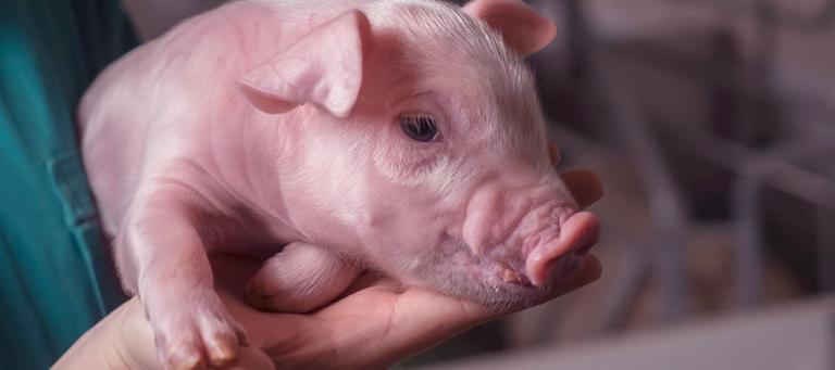
In the case of piglets, we have to know the number of affected litters, the number of piglets affected in each litter and the parity of the sows presenting with this picture.

FIGURE 1: Summary of the location and lesions of the principal causative agents in gastrointestinal outbreaks.
3. Choose the correct laboratory tests.
The joint analysis of the results of numerous laboratory techniques enables us to make a definitive diagnosis and to decide on the most appropriate course of action for the farm concerned:
Histopathology:
- For this, the samples taken are segments 3-5 cm in length. If we suspect a generalized gastrointestinal problem, consideration is given to taking samples in different portions of the large intestine, small intestine, kidney, spleen and lymph nodes. It is important to point out that, in the case of organs, the sample should not be larger than 1 cm2 in order to allow the formalin to penetrate correctly.
- The intestine is then opened longitudinally to expose the lumen and the tissue sample is carefully washed with water.
- Finally, the tissues are placed in a vial with 10% formalin.
Bacteriology:
- In this case, avoiding any animals that have been given parenteral antibiotic treatment, double ligation of the intestine is performed in the most affected areas, separating the two loops from each other by 8-10 cm.
- The segment demarcated by the ligatures is then cut and sent under strict refrigeration conditions to the diagnostic centre, never frozen.
Antibiotic susceptibility testing:
- Determination of the MIC makes it possible to establish the lowest concentration of a specific antibiotic that is able to prevent the multiplication of the bacterium that has been isolated. This test allows us to choose the antibiotic that will be most effective in treating each particular case.
Molecular (PCR):
- In this case, the procedure is similar to that when samples are taken for bacteriology, the only difference being that the sample can be frozen before dispatch.

TABLE 1: Correlation between sample taking and laboratory analysis requested
To sum up…
Managing the process, being clear about the sampling procedure and organizing the correct dispatch of the samples to the laboratory are key to the success of diagnosis in the case of gastrointestinal disorders.

FIGURE 2: Flow chart for taking samples in the case of gastrointestinal disorders in pigs
REFERENCES:
1. Ramis G., Carrasco, L., Pallarés, F.J., Astorga, R.J., Muñoz, A., Gomez, J. (2011). Patologías digestivas porcinas. Servet Editorial. pp 49-62.
2. Segalés, J., Martínez, J., Castellà, J., Darwich, L., Domingo, M., Mateu, E., Martín, M., Sibila, M. (2013) Manual de diagnóstico laboratorial porcino. Editorial Servet, pp 63-71.
Produits apparentés

Le premier vaccin intradermique sans aiguille contre Mycoplasma hyopneumoniae et PCV2, le tout en un, en suspension injectable

Vaccin vivant contre le syndrome dysgénésique et respiratoire du porc (SDRP), en lyophilisat pour solution injectable

Vérotoxine 2e recombinante purifiée contre la maladie de l’œdème en suspension injectable

Vaccin inactivé contre la rhinite atrophique porcine progressive et non progressive, en suspension injectable
Vaccin inactivé contre la colibacillose néonatale et les infections à Clostridium chez le porc, en suspension injectable

Vaccin inactivé contre les infections à Clostridioides difficile et Clostridium perfringens type A chez le porc, en suspension injectable.
Vaccin inactivé contre la parvovirose porcine et le rouget du porc, en suspension injectable
Vaccin inactivé contre le rouget du porc et la parvovirose et la leptospirose porcines, en suspension injectable

Vaccin vivant contre la souche gE négative de la maladie d’Aujeszky, en lyophilisat pour solution injectable

Vaccin inactivé contre le rouget du porc, en suspension injectable

Vaccin inactivé contre la parvovirose porcine, en suspension pour injection

Vaccin inactivé contre la pneumonie enzootique, en suspension injectable
Services apparentés

Smart Vaccination d’HIPRA est un concept révolutionnaire qui inclut un vaccin intelligent dont l’étiquette est équipée de la technologie RFID, un appareil médical assurant une vaccination précise et efficace et un nouveau monde de solutions numériques sur HIPRAlink. Tous ces éléments sont développés en interne par HIPRA.

Chez le porc, le concept de Smart Vaccination d’HIPRA est intégré dans notre vaccin novateur, l’appareil médical Hipradermic® et l’application vétérinaire professionnelle HIPRAlink®:
An innovative service based on data analysis that facilitates decision-making in swine

HIPRA Stats est un service d’analyse des données conçu pour les éleveurs.
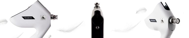
Hipradermic® est un appareil d’injection sans aiguille avancé, doté d’une connectivité sans fil pour la vaccination intradermique des porcs

L’Université HIPRA propose des programmes de formation de haute qualité dans les domaines d’intérêt stratégique pour les professionnels
Accédez à un service de diagnostic avancé assurant un maximum de confort et de fiabilité