AETIOLOGY:
Influenza viruses are members of the family Orthomyxoviridae that includes seven genera: Influenzavirus A, Influenzavirus B, Influenzavirus C, Influenzavirus D.
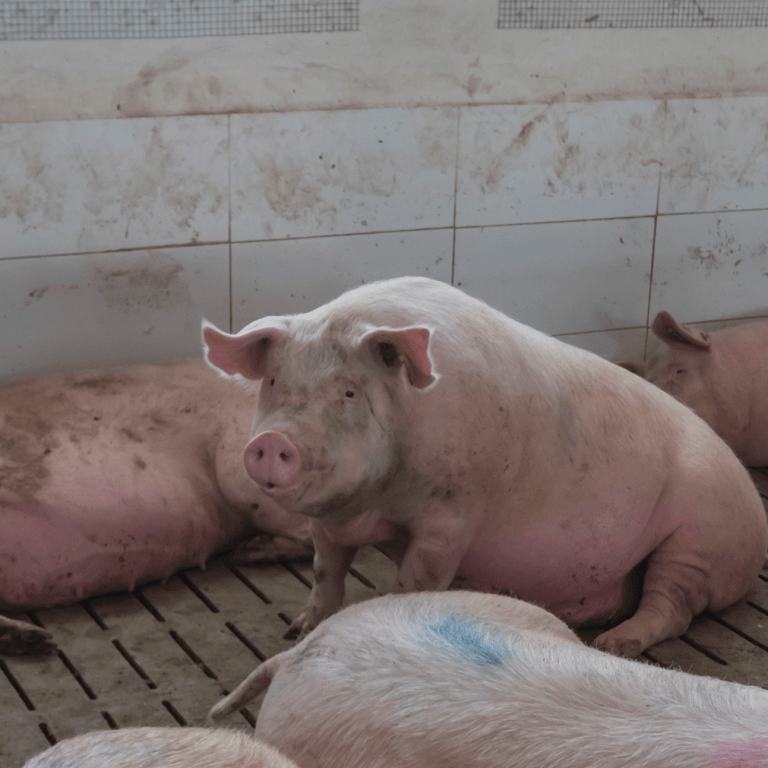
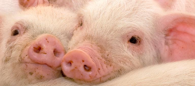
CLINICAL SIGNS:
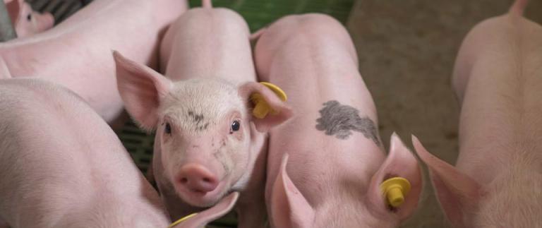
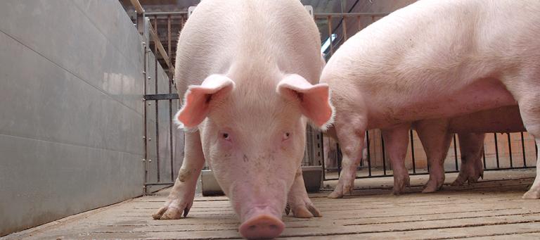
The main clinical signs are: depression, anorexia, fever (up to 42°C), prostration, coughing, dyspnea, weakness nasal and ocular mucous discharge. Mortality is generally 1%–4%. The overt course of the disease is usually 3–7 days in uncomplicated infections, with clinical recovery of the herd almost as sudden as the onset.
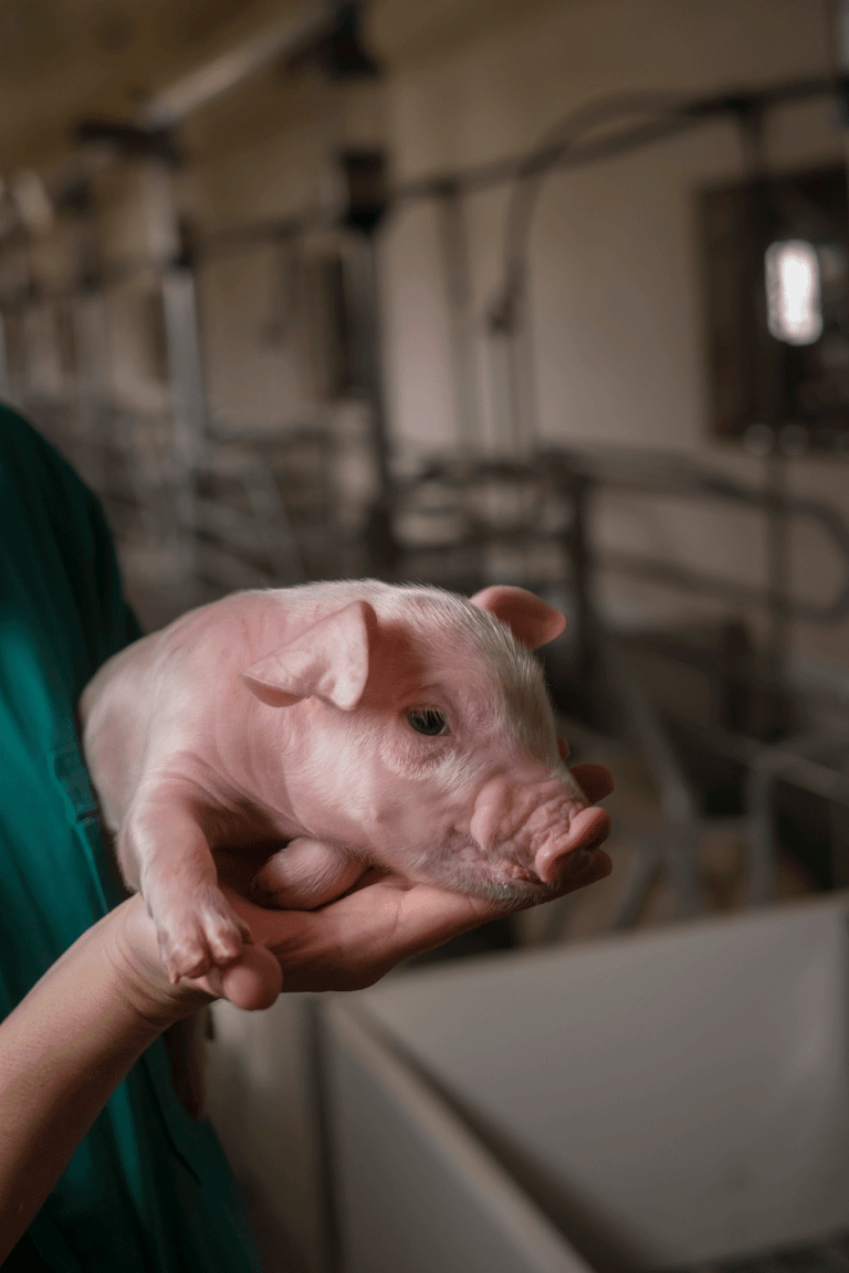
Some increase in piglet mortality has been reported, and effects on herd fertility, including abortions in late pregnancy and embryonic resorptions, may follow outbreaks in naive herds. In many herds the main economic loss is from coinfections or secondary infections.
TRANSMISSION:
A classic acute outbreak of IAV is characterized by sudden onset and rapid spread through the entire herd, often within 1–3 days. IAV cycles among pigs when clinical signs are suppressed by immune responses because pigs may become reinfected with similar or distinct strains.
LESIONS:
Lesions are confined to the thoracic cavity: affected areas of the lung are clearly demarcated, collapsed, and purplish red on gross inspection. They may be distributed throughout the lungs but tend to be more extensive and confluent ventrally. The airways contain a copious mucopurulent exudate, and the bronchial and mediastinal lymph nodes are edematous but rarely congested. There may be severe pulmonary edema, especially of interlobular septae, or a serous or serofibrinous pleuritis. Histologically the lesions are an exudative bronchiolitis with some interstitial pneumonia.
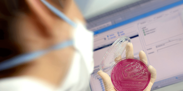
DIAGNOSIS:
Diagnosis based on clinical signs consists on observing the sudden onset of a large number of pigs showing coughing, fever, and nasal secretions. However, subclinical and chronic influenza infections are common, and in those cases, cough and nasal secretions may be sporadic.
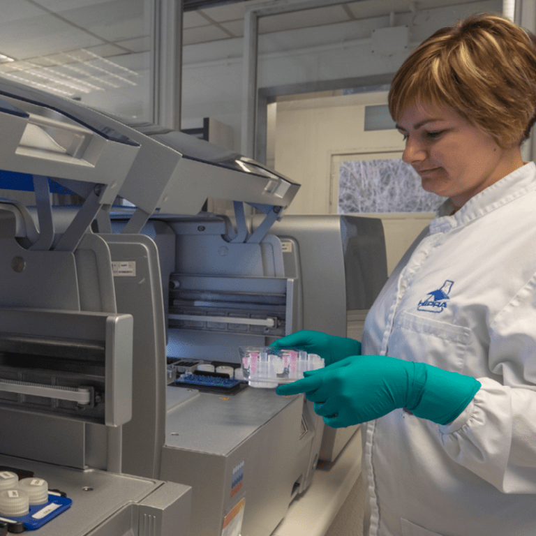
IAV is confirmed by detecting the influenza virus by RT-PCR, virus isolation, and by detecting antibodies against IAV in nonvaccinated animals. Virus can be isolated from nasal and oral secretions in the febrile phase, from affected lung tissue in the early acute stage, or from udder wipes collected from sows with infected suckling piglets. Sequencing may be needed to select epidemiologically relevant strains that may be needed for evaluating virus-specific antibodies or custom vaccine production.
TREATMENT, PREVENTION AND CONTROL:
Antimicrobials may reduce secondary bacterial infections and antipyretics may provide symptomatic relief. Vaccination and strict import controls are the only specific preventive measures. Sow vaccination attempts to maximize the transfer of maternal immunity to the progeny. Piglet vaccination is possible but interaction with maternal antibodies should be considered.
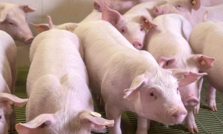
Good management practices, such as strict all-in/all-out procedures; limiting movement of pigs and sows within farrowing rooms and between pens, rooms and barns; and freedom from stress help reduce transmission and losses.