AETIOLOGY:
PRRSV type 1 (PRRSV) and PRRSV type 2 (PRRSV2) are currently regarded as two species classified with 15 other species of primate, rodent, and equine viruses in the family Arteriviridae.
CLINICAL SIGNS:
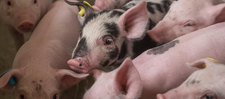
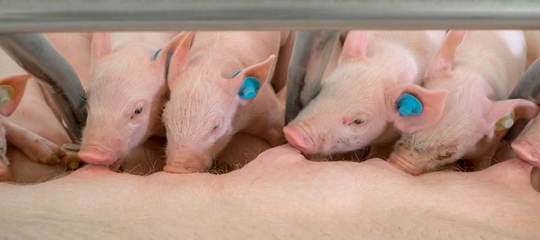
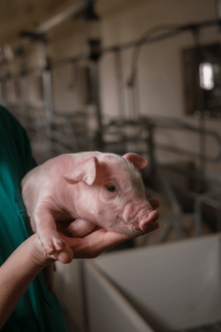
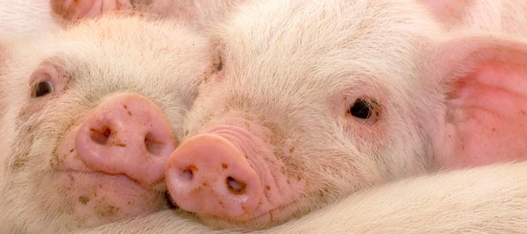
Gilts, sows, and boars: anorexia, fever, lethargy, depression and perhaps respiratory distress or vomiting. Reproductive problems, often the most obvious signs, include a decrease in the number of dams that conceive or farrow. There is usually an increase in premature farrows, late term abortions, stillborn or weak piglets and mummified foetuses. Preweaning mortality is high.
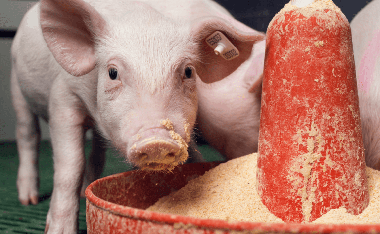
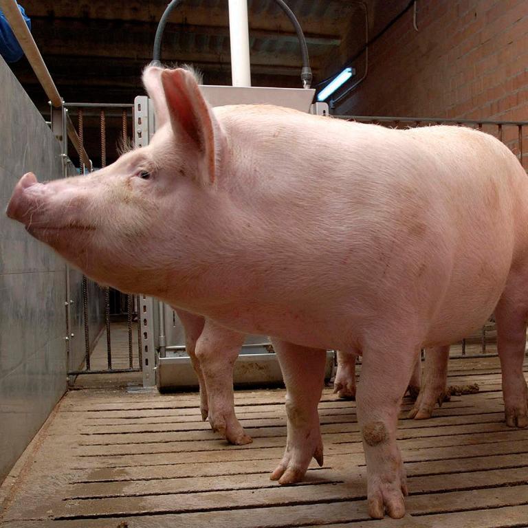
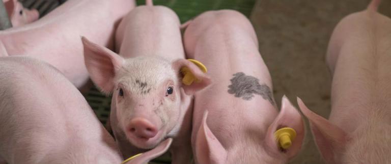
Young, growing and finishing pigs: fever, depression, lethargy, stunting due to systemic disease, and pneumonia. Sneezing, fever and lethargy are followed by expiratory dyspnoea and stunting. Peak age for respiratory disease is four to ten weeks. Postweaning mortality often is markedly increased, especially with more virulent strains and the occurrence of ever-present concurrent and secondary infections.
TRANSMISSION:
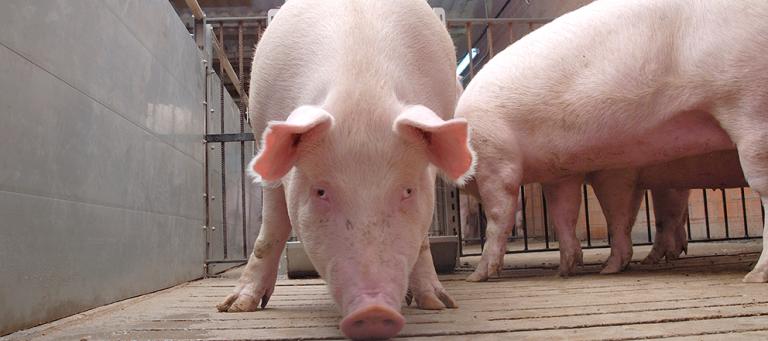
Direct transmission most commonly occurs by close contact between pigs or by exposure to contaminated body fluids (semen, virus-contaminated blood, secretions, contaminated needles, coveralls, and boots). Indirect transmission of PRRS virus has been demonstrated by several routes such as vector (personnel, trucks, fomites, feral swine, insects and rodents) and airborne transmission.
LESIONS:
PRRS virus infection generally results in mild to severe lesions in lungs and lymph nodes. The interstitial pneumonia varies from multifocal to lobular to diffuse in distribution. Lungs appear mottled and tan but are highly variable in extent. Lymph nodes are generally swollen, tan and edematous or cystic.
Foetuses and stillborn pigs some may have umbilical cord arteritis and haemorrhage; patchy distribution of slightly firm lungs (interstitial pneumonia); enlargement of lymph nodes; haemorrhages in the skin; oedema of the eyelids, periorbital tissues, colonic mesentery and various body cavities; and dehydration with prominence of the vertebral column. Sows with acute PRRS virus infection have typical lung and systemic lesions. Endometritis, myometritis and placental lesions have been reported.
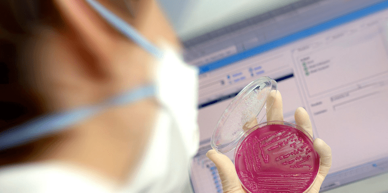
DIAGNOSIS:
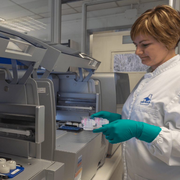
Any tentative clinical diagnosis should be confirmed by detection of the PRRS virus. This can be by virus isolation (VI), detection of PRRS antigen by fluorescent antibody tests (FAT) or immunohistochemistry (IHC), or detection of PRRS virus genome by polymerase chain reaction (PCR) and be coupled with presence of typical lesions. Sequencing is a complementary diagnostic tool for epidemiological purposes and to take decisions about measures to control the disease. Serology provides indirect evidence of infection but does not determine if there is actual disease caused by PRRS virus.
TREATMENT, PREVENTION AND CONTROL:
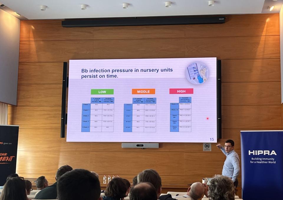
The success of the disease control depends on the combination of the following strategies: diagnostics to confirm the disease but also to understand the movement of the virus within the farm. Biosecurity consists of containment measures which prevent the entrance of a new strains as well as all internal biosecurity measures that enable us to minimise the circulation of the virus. Protocols for acclimatization and introduction of replacement animals are one of the most important points in the PRRS control. Vaccination of the animals is vital in order to minimise the clinical presentation of the disease, as it enables us to reduce the viral excretions and decrease infection pressure and the circulation of the virus on farms.