Clostridial infections in ruminants
Резюме болезней
Овцы и козы
AETIOLOGY:
Clostridia (Clostridium) are anaerobic, sporulating, gram-positive bacteria that produce toxins. They are able to survive on grasslands and meadows for a long time under extreme temperature and humidity conditions. They can be benign inhabitants of the intestine, where they live in balance with the intestinal flora, until an imbalance promotes their development and the release of toxins.
They are responsible for non-communicable infectious diseases that, from a pathogenic point of view, are usually grouped into:
- Neurotropic toxic infections: Cl. tetani and Cl. botulinum. The latter is almost non-existent in many countries.
- Enterotoxemias: Cl. perfringens and, rarely, Cl. sordellii and Cl. septicum
- Histolytic infections: Cl. chauvoei, Cl. novyi, Cl. septicum, and others.
The most common of these are Cl. perfringens infections. Type-D is more common in adults and produces enterotoxaemia, while type-C is more common in suckling lambs, resulting in necrotic enteritis, and type-B is more common in calves. The release of toxins (α, β, γ, ε, ι), with a significant haemolytic, necrotizing action is the direct cause of the clinical picture.
TRANSMISSION:
- Neurotropic:
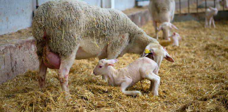
The entry of Cl. Tetani into the body usually occurs through wounds deep enough to create the anaerobic environment necessary for multiplication of the bacteria and for toxin production. Castrations, tail docking or the placement of ear tags are critical times for the animals. In the case of Cl. Botulinum, this enters the body through the ingestion of bones, grains or grass contaminated with the toxin.
- Enterotoxaemia:
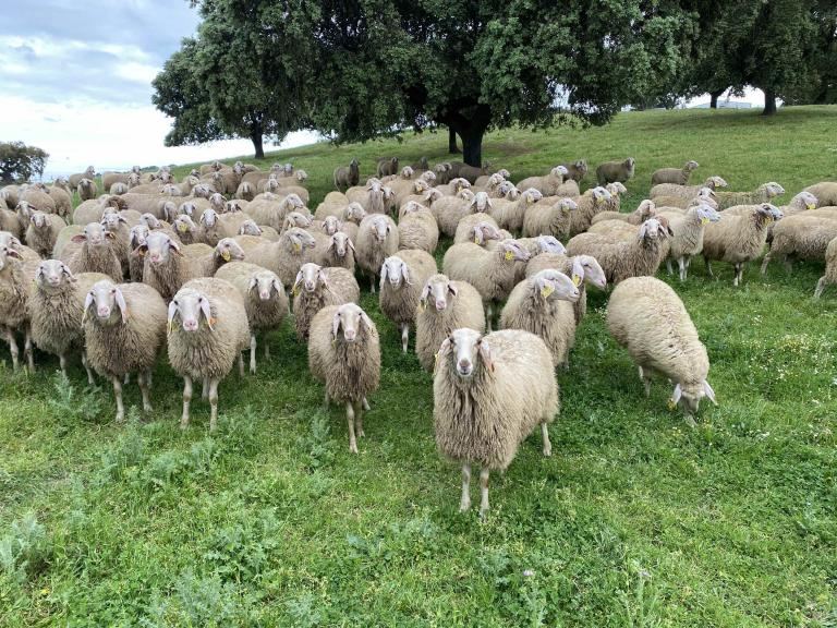
Indigestion is a clear predisposing factor in the etiopathogenesis of this process in Cl. perfringens, and may originate in groups of animals on a highly nutritional diet (excess of readily fermentable carbohydrates), in animals subjected to sudden changes in supply (change of pasture, sudden introduction of feed concentrates, etc.).
Stressful processes may also induce indigestion: transportation of animals, climate change, poor quality of pastures, etc. ultimately lead to intestinal dysbiosis, which favours the development of the disease.
- Histolytic:
Gas gangrene (Cl. septicum, Cl. novyi, Cl. sordellii, etc.) affects sheep and its route of entry is the same as that of Cl. tetani, i.e. deep wounds that can produce anaerobic conditions. It affects muscle tissue.
Blackleg (Cl. chauvoei) is an endogenous infection of calves. The bacteria is present in the musculature: in the highest growth phase of the animal, its musculature has a low level of oxygenation, thus allowing the clostridium to multiply.
Clostridia (Cl. novyi, Cl. haemolyticum) may also lead to bacillary haemoglobinuria in bovines or infectious necrotic hepatitis in sheep, respectively, both of which are closely associated to parasitic co-infection (usually Fasciola hepatica), which creates the anaerobic conditions necessary for the development and production of clostridial toxins. It may also be associated with metabolic disorders.
CLINICAL SIGNS:
- Neurotropic:
In the case of infection with Cl. tetani, spastic paralysis or convulsions may be observed, while in Cl. botulinum infections, the paralysis is flaccid.
- Enterotoxaemia:
Typically the presentation of the disease is hyperacute, first presenting signs of bloating (a result of indigestion) and often evolves into an intense and rapidly-developing febrile syndrome, combined with apathy and prostration. The animals fall over on their side with opisthotonos (their neck is stiff and turned backwards). They have difficulty breathing, and foam and exudates are present in their airways. These animals eventually die. Subacute forms of enterotoxaemia take the form of mild enteritis, where toxaemic action is low and there are few or no symptoms present. These animals recover within 3-4 days.
- Histolytic:
The most typical signs of gas gangrene are oedema and subcutaneous emphysema, along with a reddish discoloration of the skin, fever, prostration and acute death.
Calves infected with blackleg present with decay and swelling of the affected areas, fever and acute death.
In both bovine bacillary haemoglobinuria and ovine infectious necrotic hepatitis, the urine appears blood stained, there is a visible ocular icterus and sometimes blood is present in the stools.
LESIONS:
- Neurotropic:
In clostridial infections, it is difficult to find characteristic lesions at necropsy. In the case of Cl. tetani, the entry of the clostridium may be evident but nothing more.
- Enterotoxaemia:
A very rapid decomposition of the body and bloating are observed at necropsy, which are common to all clostridial processes. Exudates are also usually present in all cavities, such as pericardial, pleural and abdominal cavities. Pulmonary and mucus congestion with foam present in the airways, pericardial bleeding, hepatomegaly with parenchymal degeneration, full gallbladder. The renal parenchyma degenerates in such a clear way that this feature gives the disease its name: Cl. perfringens type D, or pulpy kidney. Other findings include abomasitis and haemorrhagic enteritis, mainly localised in the last sections of the small intestine and the first sections of the large intestine. The mesenteric lymph nodes present with hypertrophy and oedema.
- Histolytic:
Gas gangrene is characterised by a liquid or gelatinous subcutaneous exudate, with necrosis and gangrene of muscle tissues. In general, it is common to find petechiae on the serosa and fluid in cavities.
Lesions observed at necropsy in an animal infected with blackleg are very similar to those occurring in gas gangrene.
For bovine bacillary haemoglobinuria, the presence of necrotic foci in the liver, typically with a jagged edge and up to 10 cm deep, are considered pathognomonic. Other signs include icterus, petechiae on the serosa and bloody fluid in cavities. As for ovine infectious necrotic hepatitis, infected animals present with widespread congestion, focal necrosis in the liver, haemorrhagic subcutaneous oedema, icterus and bloody fluid in cavities. The presence of liver parasites is very common in both.
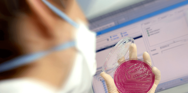
DIAGNOSIS:
They are difficult to culture due to their anaerobic condition. In most cases, simple isolation of the bacteria does not confirm the diagnosis (as they are part of the normal flora of the animal). It will be the detection of toxin that provides us with a diagnosis.
- Neurotropic:
Clinical picture of nervous system compatible with the disease, in which case other possible food intoxications (Cl. botulinum) or associated with deep wounds (Cl. tetani) are discarded. Isolating the toxin is not normally easy.
- Enterotoxaemia:
The clinical presentation is usually very acute. The type of animal infected and the necropsy (pulpy kidney) are highly suggestive of enterotoxaemia type D. The bacteria sample and the identification of toxins confirm the diagnosis. Clostridium is not usually found outside the digestive tract until a few hours after death, so only toxins are identified. A few hours later, large amounts of Clostridium invade all organs.
- Histolytic:
Existence of alterations in the musculature, with necrosis in the case of blackleg and gas gangrene and parasites in the liver as well as liver lesions, jaundice and parasites in infectious hepatitis in lambs and in bacillary haemoglobinuria.
PREVENTION AND CONTROL:
Vaccination of all at-risk animals with combined clostridial vaccines that also incorporate toxoids, or inactivated toxins. Vaccination has been long shown to be the basis of the prevention and control of these pathogens. Other measures that can be taken:
- Neurotropic:
In the case of tetanus, materials must be disinfected and tagging must be done in the most hygienic manner possible.
- Enterotoxaemia:
Proper nutritional management to avoid indigestion. Incorporating buffer substances in the animals’ diet. Vaccination of pregnant adult animals 30 days before delivery is the correct protocol, both to ensure immunity of these animals at the time that their food intake increases (concentrates in the pre-partum period), and in order to ensure successful passive transfer of immunity in newborn lambs and thus prevent dysentery.
- Histolytic:
In the case of gas gangrene, the shearing process and hygiene conditions in general should be improved. As for liver diseases caused by Cl. novyi, hepatic parasitism must be kept in check and diet should be managed well to prevent metabolic imbalances.
Сопутствующая продукция

Инактивированная вакцина против инфекций Chlamydia abortus и Salmonella abortusovis у овец, в виде суспензии для инъекций.

Инактивированная вакцина против мастита коз и овец, эмульсия для инъекций

Инактивированная вакцина против копытной гнили овец, суспензия для инъекций

Инактивированная вакцина против клостридиальных инфекций КРС, суспензия для инъекций

Инактивированная вакцина против энтеротоксемии, внезапной смерти, фомоза и инфекционного некротического гепатита, суспензия для инъекций

Инактивированная вакцина против контагиозной агалактии, эмульсия для инъекций

Инактивированная вакцина против пастереллеза жвачных, суспензия для инъекций
Сопутствующие услуги
An innovative service based on data analysis that facilitates decision-making in small ruminants.

HIPRA Stats — это сервис анализа данных для животноводческого производства.

HIPRA - университет предлагает высококачественные программы обучения специалистов

Компания HIPRA специально разработала несколько online-инструментов, помогающих ветеринарам и другим работникам сферы охраны здоровья животных упростить повседневные задачи
Быстрая и надежная лабораторная диагностика, а так же консультирование.