移至主內容
post mortem technique
Mannheimia haemolytica: from harmless commensal to dangerous pathogen
 

Bovine respiratory microbiome  

The bovine respiratory tract normally contains a mixture of bacterial colonies and various viral organisms, which can exist without causing any pathology. This community of pathogenic, symbiotic and commensal organisms is called the microbiome and it is an important dynamic and complex system that maintains  immune homeostasis in cattle.  

Colonisation of the bovine  respiratory tract starts immediately after birth and evolves quickly up to the second week of life. The nasopharyngeal microbiota of calves is dominated by Proteobacteria (Gram-negative), with copious amounts of Mannheimia, Moraxella, Bacteroides, Streptococcus and also Pseudomonas.   

The composition of the respiratory microbiota is associated with the health status of the animal.  Disease-predisposing factors often disrupt the respiratory microbial ecosystem, provoking atypical colonisation patterns and progressive dysbiosis. 

Shift from commensal to pathogen 

M. haemolytica can be a good example of dynamic change in the microbiota composition in the case of an immune homeostasis disorder 2. In healthy cattle the most commonly isolated M. haemolytica is serotype A2. This serotype does not show destructive behaviour 3 and is not usually detected in affected lung tissue, in contrast to serotype A1, which is far more aggressive. When stressors occur and immunity is compromised, A1 colonises the nasopharyngeal mucosa and quickly replaces other serotypes of Mannheimia. This shift can be regarded as the onset of pasteurellosis.  

Once a high level of A1 is established in the nasopharyngeal mucosa,  inhaled air loaded with small droplets (1-5 µm) containing A1 can reach the alveoli without any contact with the tracheobronchial defences (mucociliary escalator and TAB - Tracheal Antimicrobial Peptides), to which Pasteurellas are highly sensitive.  

The presence of Mannheimia A1 on the lung surface initiates an inflammatory reaction by interacting with  macrophages that serve as a primary line of host defence. Bronchi, bronchioles and alveoli are then subsequently infiltrated by neutrophils.

Detect and protect Mannheimia haemolytica

M. haemolytica virulence factors 

One of most important factors of M. haemolytica virulence is the leukotoxin (Lkt) produced during the logarithmic-phase of bacterial growth. Lkt production is  inversely correlated with the oxygen concentration in pulmonary tissue.  

Lkt induces dose-related4 reactions and causes changes in bovine leukocytes. At low concentrations, leukotoxin stimulates the cellular immune response through interaction with macrophages, neutrophils and lymphocytes. In higher concentrations, Lkt induces cellular osmotic swelling, pore formation in the membrane structure 4, and cell necrosis combined with a massive  release of proinflammatory cytokines and enzymes5, which exacerbate the devastating effect on the cells of the bovine immune system. Finally, a breakdown of pulmonary immunity occurs.  Lung damage is, in this case, the result of interaction between bacteria and host defences6.

Mannheimia is capable of quickly spreading the inflammatory process from the primary locus of infection to surrounding lobules4. This occurs due to a lipopolysaccharide (LPS), which has a harmful effect on the integrity of the vascular system and also increases the Lkt cytolytic activity.  

As a result, fibrinous pneumonia or pleuropneumonia develop. Lesions in the lungs appear bilaterally, mostly in cranioventral lobes. The affected tissue is usually firm, and with consolidation.  Characteristic yellowish oedema with coagulated fibrin in intralobular septa can often be spotted7. Coagulated fibrin may be also visible in intralobular lymphatic vessels. Lobes usually have a marbled appearance, ranging from pink to an even red-grey colour. Dark pink foci are usually surrounded by a thin brighter line, containing an accumulation of inflammatory cells which creates an easy-to-spot, sharp demarcation.  Coagulated fibrin is also present inside the bronchi. In advanced stages of mannheimiosis, these changes can be visible in the middle and caudal lobes. In the case of fibrinous pleuropneumonia, the pathology image is similar but fibrinous pleuritis is more intensive.

Prevention of Mannheimiosis 

In the past, prevention of bovine mannheimiosis was based almost exclusively on systemic antibiotic therapy. Prophylactic administration of long-acting antibiotics in calves prior to stressful events had become common practice, but today's healthcare professionals and farmers know that antibiotics are a double-edged sword. Current evidence indicates that the widespread use of antibiotics contributed to the emergence of multiple antibiotic-resistant strains of M. haemolytica8

This is why other non-antibiotic approaches to bovine mannheimiosis such as vaccination are  constantly being researched.  

Since the 1980s, vaccination with whole inactivated bacterins has been performed with questionable efficacy. Killed bacterins of course induce a rise in agglutinating antibodies but they have almost no antitoxic response to the most important virulence factor- leukotoxin.  In fact, there is evidence that bacterin-vaccinated animals can be more susceptible to disease9,10.  Better results were observed after immunisation with live attenuated bacteria, but for reasons of safety they are no longer commonly used in Europe.  This is why researchers' efforts have focused on developing leukotoxin-based vaccines11. This type of immunisation stimulates a rise in both antitoxin and opsonising antibodies, providing superb immunity with a 50-70% reduction of clinical signs and lung lesions.  High efficacy of anti-leukotoxin vaccines  is also a result of confirmed, universal cross-protection between different Lkt-types produced by serotypes other than the most common A1 (A5,A6,A8,A9 or A12)12

Conclusion 

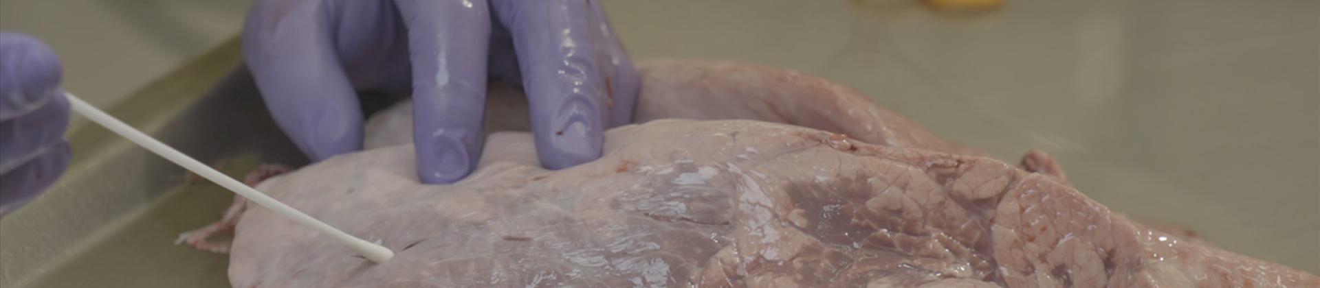
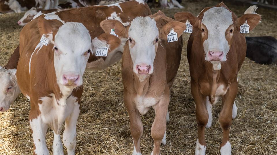
Mannheimia heamolityca is typically associated with fatal pneumonia13. Calves usually become infected immediately after birth,  due to nasal contact with their mothers. In healthy animals, Mannheimia behaves as a commensal, until stressful factors appear.  BRD prevention should not be based on regular antibiotic use but on welfare and management prevention practices, such as vaccination, in order to control this costly disease in cattle.  


Author: Wojciech Ptak, DVM .


References: 

1. C. Blakebrough-Hall, A. Dona, M. J. D’occhio, J. McMeniman & L. A González, 2020. Diagnosis of Bovine Respiratory Disease in feedlot cattle using blood 1H NMR metabolomics. Sci Rep 10:115 

2. Edouard Timsit, DVM, PhDa, Chris McMullen, BScb, Samat Amat, MSc, PhDb,c,d, , Trevor W. Alexander, PhDc, 2020. Respiratory Bacterial Microbiota in Cattle From Development to Modulation to Enhance Respiratory Health Vet Clin North Am Food Anim 36, 297–320. 

3. Griffin, D., Chengappa, M.M., Kuszak, J., McVey, D.S., 2010. Bacterial pathogens of the bovine respiratory disease complex. Vet Clin North Am Food Anim Pract 26, 381-394. 

4. Roger J. Panciera, Anthony W. Confer. Pathogenesis and Pathology of Bovine  Pneumonia Vet Clin North Am Food Anim Pract. 2010 Jul; 26(2): 191–214. 

5. Jeyaseelan, S., Sreevatsan, S., Maheswaran, S.K., 2002. Role of Mannheimia haemolytica leukotoxin in the pathogenesis of bovine pneumonic pasteurellosis. Anim Health Res Rev 3, 69-82. 

6. Mosier, D., 2015. Review of BRD pathogenesis: The old and the new. Animal Health Research Reviews, 15(2), 166-168.  

7. Malazdrewich C, Thumbikat P, Maheswaran SK, 2004. Protective effect of dexamethasone in experimental bovine pneumonic mannheimiosis. Microbial Pathogenesis, 36(4), 227-236.  

8. Zecchinon, L., Fett, T., Desmecht, D., 2005. How Mannheimia haemolytica defeats host defence through a kiss of death mechanism. Vet Res 36, 133-156. 

9. Auad J, Carbonero Martínez A, Victoria Maure MV, Daffner JF, Gleser HD, 2001. Inoculation trial and humoral response with an inactivated oily vaccine against the bovine respiratory complex (BRC). Veterinaria Argentina, 18(176), 430-439. 

10. Stevens RD, Dinsmore RP, Ellis RP, Katsampas M, 1997. Morbidity and mortality in young Holstein heifer calves vaccinated with a P. haemolytica leukotoxoid. Large Animal Practice, 18(6):23-29. 

11. Tobias Oppermann, Nadine Busse,  Peter Czermak, 2017.  Mannheimia haemolytica growth and leukotoxin production for vaccine manufacturing — A bioprocess review. Electronic Journal of Biotechnology, 28, 95-100. 

12. Rice, J. A., Carrasco-Medina, L., Hodgins, D. C., Shewen, P. E., 2007. Mannheimia haemolytica and bovine respiratory disease. Animal Health Research Reviews, 8(2), 117-128.  

13. Laëtitia Dorso, Maud Rouault, Claire Barbotin, Christophe Chartier, Sébastien Assié, 2021. Infectious Bovine Respiratory Diseases in Adult Cattle: An Extensive Necropsic and Etiological Study. Animals (Basel). 2021 Aug; 11(8): 2280.