移至主內容
10 Step Post Mortem Technique
A respiratory postmortem in 10 steps
 

Necropsy procedure for the diagnosis of the respiratory disease syndrome

One of the most common collective diseases in cattle is the respiratory disease syndrome. It usually takes the form of epidemic outbreaks, mainly affecting young animals, with highly variable morbidity and mortality. It causes significant losses, both on farms where cattle are fattened and on those where they are bred and reared. It is a process with an infectious aetiology, although its appearance can depend on the existence of environmental or management factors that predispose the animals to it. The pathogenic agents involved are mainly viruses and bacteria.

The technique of express or targeted necropsy described below is recommended in two scenarios:


  • Whenever there is a prior and unequivocal clinical diagnosis of respiratory disease syndrome and we are certain that the losses are caused as a consequence of this. In this situation, the objective will be not so much to determine the cause of death, which we already know, but to try to identify the microorganisms involved in the outbreak.


  • When we are faced with dead animals and we have no clinical data on the process, but we observe predisposing factors that lead us to suspect that it may be a respiratory disease. These are primarily unfavourable housing conditions, individuals of different origins grouped together, poor management and other causes of stress. We should also take the history of the disease on the farm into account.


If this rapid technique does not reveal conclusive lesions and as long as we wish to carry out a more thorough examination, we can continue by examining other cavities and completing the necropsy with an inspection of all the tracts and systems.

The macroscopic study of the lesions will provide us with immediate information that we should add to our clinical and epidemiological observations, establishing a hypothesis on the aetiology of the case. Taking samples for subsequent analysis in the laboratory may confirm suspicions or reveal the presence of other microorganisms. In the necropsy procedure detailed below, we will obtain samples that can be used for processing by the Polymerase Chain Reaction (PCR) technique that will determine the presence or absence of the most common pathogens. When the samples are sent to the laboratory, a panel should be selected that includes the most relevant respiratory pathogens: viruses such as the BRSV, IBR, BVD or Pi3 and bacteria such as M. haemolytica, H. somni, P. multocida or M. bovis. Sample taking will be slightly different (aseptic) if culturing is the intention; it should be borne in mind that cultures may limit the diagnosis of bacteria such as H. somni or M. bovis and are not applicable in the case of viruses.

In essence, the aim of the technique that is proposed here is to enable the veterinarian faced with an outbreak of respiratory disease syndrome, or on farms with endemic problems, to carry out a rapid and simple necropsy in the field without the need for specific training or extensive previous experience, using only a basic instrument set. The simplification of the procedure and the reduction in the time taken will in no way affect the scientific rigour with which the veterinarian should approach a necropsy. It merely involves an adaptation to the clinical working situation, allowing it to be carried out as often as necessary.

In this article, we limit ourselves to describing the technique. The assessment of the lesions and the interpretation of the laboratory results are subjects that will be dealt with in subsequent chapters.

Instrument set and basic equipment

Before starting, we must ensure that we have available the equipment for the performance of the necropsy and for appropriate sample-taking. A sharp knife is essential; we will also need a sharpener, scissors, small axe (optional), forceps, 6 sterile swabs without medium and with plastic handles, 2 sample vials.

Detect & Protect materials

10-step technique in 10 minutes


1. Place the cadaver in the left lateral decubitus position.

The position is important in the case of ruminants, as the volume of the rumen is large and can make examination of the abdominal cavity difficult if it needs to be accessed.


2. Open the stifle joint to examine the synovial capsules.

When an incision is made into a healthy joint, the presence of synovial fluid is minimal. If synovitis is suspected, a sample of synovial fluid should be taken to investigate the possible presence of Mycoplasma bovis or Histophilus somni, respiratory pathogens that also tend to colonize these tissues.


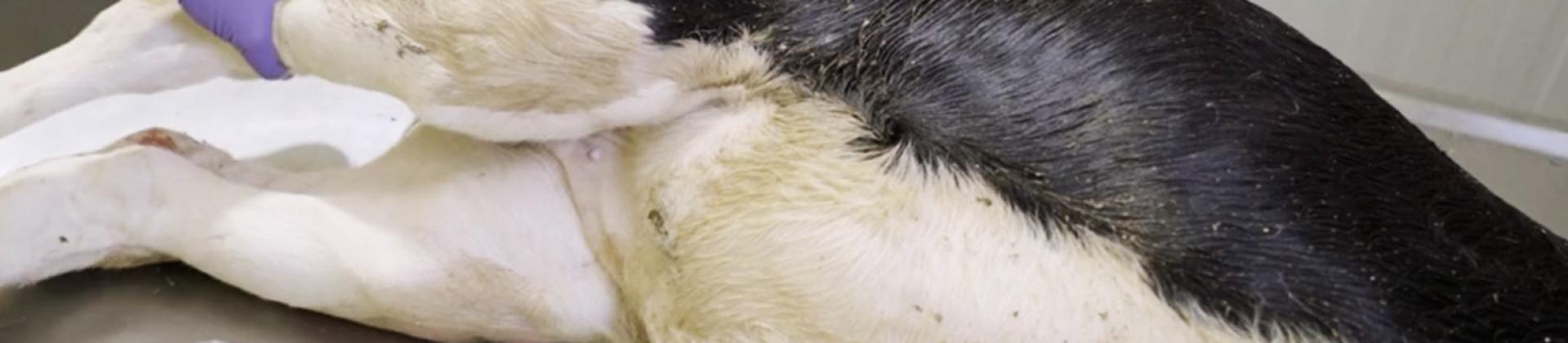
3. Dissect the right foreleg.

Make an incision in the axilla, the leg muscle attachment site, at the same time as the limb is abducted, until the thorax can be easily accessed.


4. Expose the trachea as far as the pharynx.

Detach the trachea completely, including the pharynx, so that the whole respiratory tract can be subsequently removed.


5. Open up the ribs.

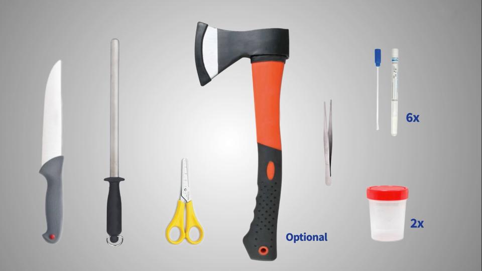
Make long incisions in the intercostal spaces, ensuring that the lung is not damaged. Cut along the line of the sternum, which will allow the thoracic cavity to be opened. In older animals, an

axe may be needed to cut along the line of the sternum. Breaking the ribs manually, open the cavity until the lung is exposed.

At this point, we are able to observe the appearance of the lung and the visceral and parietal pleura. We will also be able to see if there is any abnormal fluid in the cavity.


6. Remove the respiratory tract.

Completely remove the respiratory tract, from the pharynx to the lungs. Place it on a clean surface which allows the organs to be manipulated easily.


7. Inspect the lung tissue.

Use scissors to make several cuts deep into the lung parenchyma, along the line that separates the healthy from the unhealthy tissue. Repeat this in the affected area at a distance of 1 or 2 cm from the previous incisions. Take samples by scraping vigorously with the swabs inside each incision in order to detect the presence of viruses and bacteria. Small samples of affected tissue may be cut out and placed in a vial to be sent to the laboratory.


8. Check for the presence of abscesses or other lesions in the pharynx.


9. Open the trachea as far as the bifurcation.

Note the presence of lesions and collect samples by rubbing vigorously with a swab to look for the presence of viruses, such as the Bovine Herpesvirus I (IBR), which has a great affinity for this organ.


10. Cut the myocardium into strips.

Make several incisions to reveal the presence of infarcts, abscesses or myocarditis, indicators of possible Histophilus somni infection. If this is suspected, samples can be taken with a swab for PCR.

Conclusion

The main advantage of this necropsy technique is that it can be incorporated into working routines in the field. It can be performed whenever we are faced with a cadaver on a farm visit without the need for advance planning and without having to alter our schedule for the day, as it will take very little of our time.

Apart from the diagnostic information that it provides, it is usually very useful as it enables the farmer to have a greater awareness of the problem and of the economic consequences, encouraging the introduction of preventive measures that we are able to suggest.


Author: Ivan Mato DVM PhD. Technical Services, HIPRA (Spain)


References:

Andrews J.J, Van Alstine W.G, Schwartz K.J. (1986). A basic approach to food animal necropsy. Veterinary Clinics of North America: Food Animal Practice. 2(1): 1-29.

Griffiths I. (2005). Postmortem examination of cattle and sheep. Journal of the British Veterinary Association. 27(9): 458-465

Helman RG. (2020). The role of the veterinary diagnostic lab in the management of BRD. Anim Health Res Rev. 21(2):160-163

Mason G.L. and Madden D.J. (2007). Performing the field necropsy examination. Veterinary Clinics of North America: Food Animal Practice. 23(3): 503-526.

Nietfeld J.C. (2010). Field necropsy techniques and proper specimen submission for investigation of emerging infectious diseases of food animals. Veterinary Clinics of North America: Food Animal Practice. 26(1): 1-13.

Ruth G.R. (1986). Necropsy of adult cattle. Veterinary Clinics of North America: Food Animal Practice. 2(1): 119-127.

Wagner S. (2007). Necropsy techniques in cattle. Proceedings of the Fortieth Annual Conference. American Association of Bovine Practitioners: 203-204. September 20-22. Vancouver, British Columbia.