2d. Clostridium Novyi in Sows (Case report)
Posts
Porcino
Sudden Death Caused by Clostridium novyi in Sows in a Conventional Herd
Article written by:
Name & Lastname Escobet Riu
Position, company, Country
Sudden deaths in sows often leave piglet producers and veterinarians with a mystery. Nevertheless, the causes are rarely investigated. Only if the cause of death is known, the underlying problem can be addressed appropriately. In the case described here, an infection with Clostridium novyi (sudden death) was identified as the cause of death on a German pig farm. Since measures were implemented, no further deaths associated with this disease occurred.
Summary
This case report describes an investigation of sudden death of sows on a piglet producing farm in Germany. The sows were in the gestating unit and did not show clinical signs before. Necropsy of a pregnant gilt was performed immediately after finding. Nevertheless, the animal showed tympany.
Gross lesions were subcutaneous oedema and purple discolouration of the skin. Body cavities were filled with foul-smelling serosanguinous exudates and gas. The necrotic, discoloured liver had a honeycomb appearance due to gas bubble infiltrations (aerochocolate liver) and showed rapid decompensation.
Samples of the liver were subjected to bacteriological investigations where Clostridium novyi was detected. Histological lesions indicated peracute inflammation. Clostridium novyi was the suspected cause of death, based on the history of sudden death, necropsy and bacteriological findings.
In case of unexpected mortality in gestating sows Clostridium novyi should be considered. Necropsy and sample collection must be performed as soon as possible after death and cultural findings should be interpreted in combination with the history and gross lesions.
Introduction
Members of the genus Clostridium are obligate anaerobic, gram-positive, spore-forming bacteria. They occur ubiquitously and are a minor component of the intestinal microbiota of humans and animals [20].
Their spores are very resistant to external influences and can survive for a long time in the environment. Their invasion capability is low. They enter the body though wounds or are taken in orally.
Based on the produced exotoxins, clostridia are classified into different toxin phenotypes [5]. The major toxins include the alpha toxin and the beta toxin. They cause a change in the permeability of the cell membranes and lead to a loss of integrity of the vascular endothelium, resulting in necrosis, oedema as well as serosal effusions and peracute or acute death [5, 21].
Clostridia cause diseases in humans and various domestic animals. Based on the characteristic lesions, they are classified into neurotoxic, histotoxic and enteric clostridia. Neurotoxic clostridia include Clostridium (C.) tetani (causes tetanus) and C. botulinum (causes botulism).
Both diseases have also been described in pigs but are rare [21]. In pigs, enteric clostridial infections are of the greatest relevance. C. perfringens type A and C, as well as C. difficile, are typical pathogens of suckling piglet diarrhoea.
They mainly affect new-born suckling piglets in the first days of life. Diseases rarely occur after the first week of life. The animals show diarrhoea (yellow to grey). Frequently, gas bubbles are present. Gross lesions are found in the small intestine [21]. C. perfringens has also been detected in association with enterohaemorrhagic syndrome (EHS) [20].
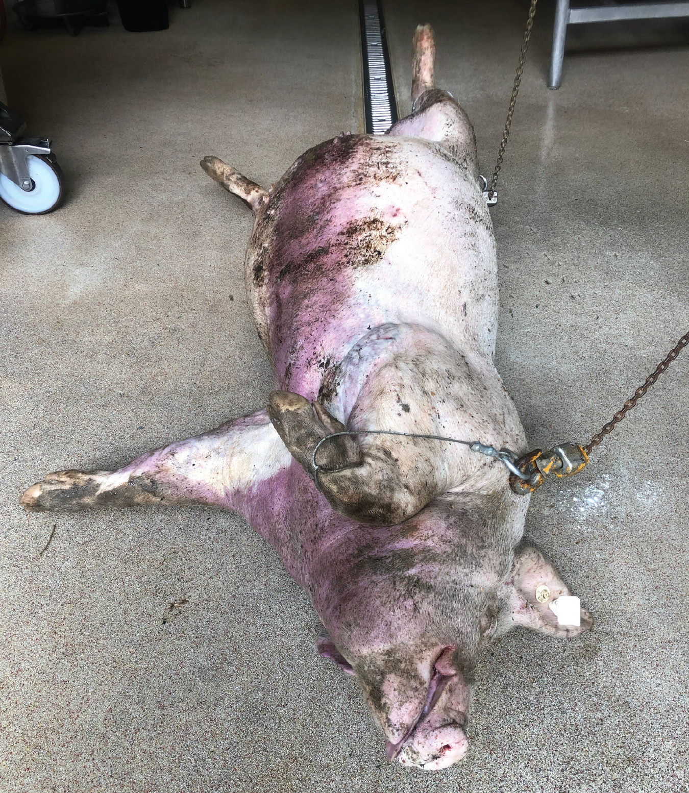
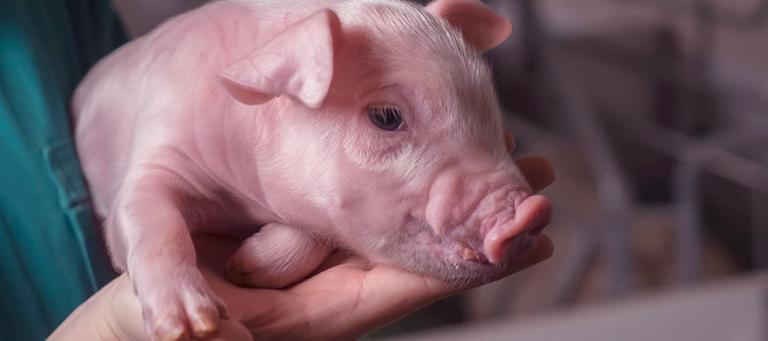
Gilt at necropsy with severe distension and purple discoloration of the skin.
Histotoxic clostridia, which cause gas oedema diseases, include C. septicum, C. chauvoei, C. sordelli, C. perfringens type A and C. noyvi. They mainly cause diseases in ruminants but are also described in pigs. Infection occurs via wounds or by spores taken in orally from the environment. Under certain conditions, the spores can become vegetative and gas gangrene develops [21].
Sudden deaths result in high economic losses for the pig farms. The immediate loss of production is caused by the dead sow and her piglets. A higher replacement rate leads to higher production costs and comes with a decreased average age of the herd. This increases the risk of disease due to the higher percentage of younger sows [23].
Sow mortality is often higher in summer than in winter, for which extreme temperatures are suspected to be responsible [4, 13]. The causes of deaths in sows are rarely investigated. Besides the economic losses of a high sow mortality, animal welfare as well as the influence on the farmer’s psyche must not be underestimated [23].
In the present case, an investigation of sudden death of sows in a conventional herd is described.
Case Description
Farm
The farm produces in a two-week production cycle with 250 sows and subsequent fattening unit. Replacement gilts are produced on site, so that only semen is sourced externally.
Directly after artificial insemination until one week before farrowing, gestating sows are kept in one large group with a demand feeding station. The sow herd is regularly mass vaccinated against PRRSV, influenza virus, parvovirus and erysipelas.
Six and three weeks before farrowing, the sows are vaccinated against rhinitis atrophicans and with an autogenous vaccine against E. Coli, Clostridium perfringens type A, Mycoplasma hyorhinis, Staphylococcus hyicus and Streptococcus suis.
Case history
At the end of August 2020, three sows had suddenly died on the farm within one week. According to the farm manager, the sows did not show clinical signs beforehand. During the inspection of the herd by the veterinarian, no abnormalities were found in the sow herd.
The deaths occurred in the gestating unit, the sows had different parities and were in different stages of pregnancy. One gilt was found dead on her 28th day of pregnancy. Immediately afterwards, she was taken for post-mortem examination to the veterinary practice “Tierärzte Hohenlohe”.
Necropsy
The body condition of the sow was good to very good. The abdominal skin was intact but showed dark purple discolouration.
On external inspection, the abdomen was grossly distended (Fig. 1). Generalised subcutaneous oedema was found. Subcutaneous gas bubbles could be felt bursting under the fingers and a crackling sound could be heard. A foul odour emerged when the carcass was opened. The pleural and peritoneal cavities were filled with reddish-brown, foul-smelling fluid and gas.
The lungs appeared well retracted with small gas bubbles on the surface. The liver was discoloured (dark red to greybrown), with necrotic areas and infiltrated by gas bubbles (Fig. 2 and 3). A sweet and foul odour of decomposition emerged from the liver. The liver parenchyma was friable
and had a spongy appearance (aerochocolate liver).
Sections of the small intestine were dark red in colour and moderately distended. No torsion of the abdominal organs could be detected. All other organs, not explicitly mentioned here, were without pathomorphological findings. Samples of liver tissue were submitted to the laboratory for bacteriological and histological examination (aCare Lab GmbH & Co. KG, Leipzig, Germany).
Laboratory results
Bacteriological examinations of the liver showed C. novyi, as well as C. perfringens and streptococci. Signs of necrosis, such as insufficient staining of the hepatocytes and karyolysis, were found in the liver tissue during pathohistological examination.
The liver parenchyma showed empty cavities of different sizes. The interstitium contained high quantities of rod-shaped bacteria, that resembled clostridia species. Lacking evidence of a prolonged inflammatory process, a peracute inflammatory process seemed most likely.
Summary and diagnosis
According to the case history, the sow had died suddenly, without any clinical signs beforehand, and was already severely distended shortly after death. Despite necropsy being performed immediately after death, a foul smell was noticeable. The body cavities were filled with foul-smelling fluid and gas.
The liver had a honeycomb appearance with gas bubbles on the surface (aerochocolate liver) and necrotic areas. Bacterial culture of C. novyi from the liver and the histological findings completed the picture.
The infection with Clostridium novyi (sudden death) as the suspected cause of death was confirmed.
Discussion
Clostridium novyi was first described by Frederick Novy in 1894 and is classified as obligate pathogenic histotoxic clostridia. Based on the exotoxins, it is categorised into four toxin types (Tab. 1) [10].
The alpha toxin causes necrotising hepatitis, severe oedema, congestion, and haemorrhage in various organs. The cell-cell connections are loosened, and the permeability of the cell membranes is altered [5].
Venous congestion leads to dark purple discoloration of the skin. The alpha toxin is produced by C. novyi type A and B and differs from those of C. septicum and C. perfringens. The beta toxin is produced by C. novyi type B in small quantities and causes intravascular haemolysis and haemorrhages [10, 20].
In humans, C. novyi occurs along with C. perfringens as the causative agent of gas gangrene [16]. This febrile, highly painful wound infection is commonly associated with war injuries and is characterised by crackling sounds under the skin. In sheep, cattle, goats and horses, C. novyi causes infectious necrotic hepatitis (Big head/Black disease) [20]. In pigs, infections with C. novyi types A and B are associated with sudden deaths [5, 20, 21].
The hallmarks are rapid and severe distension of the animals, unusually rapid decomposition, bloodstained fluids in body cavities as well as subcutaneous oedema. Particularly prominent are necrotic liver changes with gas bubbles [5]. Duran and Walton [5] describe C. novyi infection mainly in multiparous sows with good body condition around farrowing. Various authors describe the disease as rare [2, 5, 7, 21].
However, 40 % of sudden deaths in the US could be linked to C. novyi [19]. Only little data is available on the prevalence. In a study conducted in Spain, C. novyi was detected in 74.23 % of the breeding farms which were sampled [3].
The infection route for C. novyi can be exogenous from the environment, but it is also part of the large intestine microbiota in pigs and can as well be detected in liver tissue of healthy pigs [22].
For reasons unknown today, in some individuals, clostridia migrate into the liver via the bile ducts. It has not been finally clarified yet how C. novyi enters the liver and through which factors the spores are activated. In general, a low oxygen concentration triggers the germination, multiplication, and toxin production of clostridia.
The pathogenesis of C. novyi is mainly dominated by the effect of the alpha toxin, which is produced by both C. novyi type A and B [21]. It leads to liver necrosis, oedema, and serosal effusions. Acute or peracute deaths occur [21].
Diagnosis
The diagnosis of C. novyi infections is difficult, as post-mortem examination must be carried out immediately after death. It is recommended to examine the carcasses no later than twelve hours after death [6, 8]. The more time passes before samples are taken, the greater the risk of false positive results.
Sporulated C. novyi can also be detected in livers of animals that have died of other causes and can become vegetative after death. Demonstration of C. novyi as the only evidence is therefore not sufficient to confirm the assumption [17, 23].
The results of the bacteriological investigations should always be assessed in the context of the clinical history (sudden death in sows) and the gross lesions. In the case described here, the gilt did not show any clinical signs beforehand.
Immediately after death, the animal was already severely distended with subcutaneous oedema and dark purple discoloration of the skin, as described in other case reports [8]. Exudates were observed in body cavities, as also mentioned in other cases [1, 6, 7]. An enlarged spleen is reported in literature [6, 7, 8], but was not observed in the present case.
Hallmarks are an enlarged, friable, gas bubble-infiltrated liver with a honeycomb appearance (aerochocolate liver) in a well-preserved carcass [5, 6]. Since these alterations were present and there were no signs of other causes of death, the tentative diagnosis "sudden death due to C. novyi“ was made.
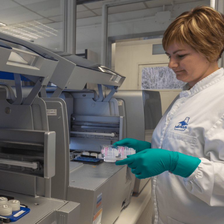
For confirmation of the hypothesis, C. novyi can be isolated from altered tissue. Detection is possible in smears of the altered organs [8, 21]. In the Gram stain, C. novyi presents as a straight, seldomly slightly curved, gram-positive rod with rounded ends and subterminal spores.
In older cultures, Clostridia may occur gram-labile or gram-negative. Cultivation is carried out on appropriate clostridial culture media under anaerobic conditions [20]. Gas chromatography or MALDI-TOF mass spectrometry can be used for species diagnosis [18, 20].
Currently, no PCR is available in Germany for the precise determination of C. novyi. In the opinion of the author, the genotypic characterization of C. novyi into type A or type B plays only a minor role in practice. Both types produce the alpha toxin, which is made responsible for the lesions, and appear in the environment [21].
The hypothesis of a C. novyi infection was confirmed by the bacteriological culture of C. novyi and the histological lesions of the liver tissue. Since samples were taken immediately after the sow was found dead, postmortem colonisation was considered unlikely.
Predisposing factors
Most case reports describe sudden death in sows in outdoor pig units [6, 7, 8, 11]. The authors discuss conditions such as weather changes, extreme temperatures, and high infection pressure from the environment as potential risk factors [2, 6, 7, 11]. Reports of C. novyi infections at conventional pig farms are rare [5, 15, 24]. Likewise, extreme climatic conditions can be difficult here as well.
In the present case, the deaths occurred in the gestating unit of a conventional farm. At that time, it was very warm in the barn due to high outside temperatures. Co-infections, such as dysentery, cystitis, metritis, and enteritis [8], as well as respiratory diseases and erysipelas are discussed as possible triggers [6].
Unlike in sheep, where liver fluke is known as possible causes for the multiplication of C. chauvoei [20], liver damage caused by parasites has not been described yet as a predisposing factor for the multiplication of C. novyi in pigs. In general, all immunosuppressive factors could however contribute to the development of the disease [8].
Two days before farrowing to three weeks after, the risk of disease seems to be higher [2, 7, 14]. Increased feed intake and stress after farrowing are discussed as potential causes [12]. García et al [8] describe C. novyi as a common cause of death in sows in the perinatal period.
They link this to the changes in the immune system, the great metabolic effort, as well as the weakening of the animals after farrowing. On the other hand, there are also reports of cases unrelated to farrowing.
For example, Jandowsky et al [11] describe a case concerning pigs of different age and gender. In the case described here, the deaths occurred in the gestating unit and the sow examined died on the 28th day of gestation.Apart from a frequent occurrence in sows with more than four litters, a good body condition is considered as a risk factor [5].
Body condition has also been described as a risk factor in sheep. Well-developed sheep seem to be more affected by necrotic hepatitis [20]. In summary, factors such as good to very good body condition, a large amount of ingested feed and late pregnancy can lead to inadequate blood supply of the liver.
This can cause changes in the liver tissue with tissue damage and anaerobic conditions. It is possible that other diseases also contribute to a reduction of oxygen in the liver tissue and thus favour the replication of C. novyi [5].
Treatment and prevention
A good health status of the herd seems to play a key role in preventing the infection [8]. Farm-specific preventive measures, regular antiparasitic treatment as well as appropriate feeding and water supply help to maintain a good level of health of the herd.
The role of co-infections on the infection process has not been conclusively clarified. Since a good to very good body condition is considered a predisposing factor, it should be regulated if necessary.
Clostridia spores occur ubiquitously and are extremely resistant to environmental influences. Carcasses should be removed as soon as possible to minimise contamination of the environment. Elimination of the pathogen from the herd seems hardly possible [8].
Some authors recommend antibiotic treatment to reduce mortality during an acute outbreak of the disease in the herd [8, 19]. Antibiotic treatment only affects clostridia.
However, it should be noted that toxins, playing the crucial role in the pathogenesis, are not influenced by antibiotics. Due to the peracute course of disease, treatment of sick animals is not possible.
It is possible to vaccinate sows against C. novyi. In Germany, two vaccines for pigs are commercially available (Suiseng®, LABORATORIOS HIPRA, S.A; Covexin® 8, Zoetis Germany GmbH).
In the event of an acute outbreak, the manufacturers recommend vaccinating the entire herd twice, at an interval of four weeks. Subsequently, sows can be vaccinated three weeks antepartum [8, 14].
Vaccination with Suiseng® has shown to induce neutralising antibodies against the alpha toxin and reduce clinical signs [9]. Jandowsky et al [11] even consider vaccination prophylaxis essential when pigs are kept outdoors.
Measures and follow-up in the current case
As the number of deaths in the described case accumulated, the sow herd was initially treated with amoxicillin to reduce the clostridial load. Simultaneously, the sow herd was vaccinated against C. novyi (Suiseng®, LABORATORIOS HIPRA, S.A).
After four weeks, at the time of the booster vaccination, only one additional sow had died. However, this occurred after a difficult farrowing and the death was not associated with C. novyi.
To permanently minimise the risk, it was recommended to continue the vaccination of the sows against C. novyi, to improve the climate in the gestating unit and to check the sows‘ body condition and adapt it if necessary.
Furthermore, the farm was advised to disinfect the water with chlorine dioxide. Here, it was important to increase the concentration of chlorine dioxide slowly over an extended period of time to avoid a sudden release of biofilm in the pipes.
In practice, the combination of water disinfection with chlorine dioxide, an initial antibiotic treatment of the animals and the simultaneous vaccination of the sows against C. novyi has proven to be a successful approach. In similar cases associated with C. novyi infections, no further deaths occurred after these measures.
Productos relacionados

La primera vacuna intradérmica sin aguja frente a Mycoplasma hyopneumoniae y PCV2, todo en uno, en suspensión inyectable

Vacuna viva frente al síndrome respiratorio y reproductivo porcino (PRRS), en liofilizado inyectable

Verotoxina 2e recombinante purificada frente a la enfermedad de los edemas, en suspensión inyectable

Vacuna inactivada frente a rinitis atrófica progresiva y no progresiva, en suspensión inyectable
Vacuna inactivada frente a colibacilosis neonatal porcina y clostridiosis porcina, en suspensión inyectable

Vacuna inactivada contra infecciones debidas a Clostridioides difficile y Clostridium perfringens tipo A en porcino, en suspensión inyectable.
Vacuna inactivada frente a parvovirosis porcina y mal rojo, en suspensión inyectable
Vacuna inactivada frente a erisipela, parvovirosis y leptospirosis porcina, en suspensión inyectable

Vacuna viva frente a la enfermedad de Aujeszky cepa gE negativa, en liofilizado inyectable

Vacuna inactivada frente al mal rojo, en suspensión inyectable

Vacuna inactivada frente a la parvovirosis porcina, en suspensión inyectable

Vacuna inactivada frente a la neumonía enzoótica, en suspensión inyectable
Servicios relacionados

Smart Vaccination de HIPRA constituye un concepto revolucionario que combina una vacuna inteligente que incorpora tecnología RFID en su etiquetado, un dispositivo de vacunación que garantiza la precisión y la eficiencia, y un nuevo mundo de soluciones digitales a través de HIPRAlink Vaccination. Todos estos elementos han sido desarrollados de forma interna por HIPRA.

En el ganado porcino, SMART VACCINATION de HIPRA incluye nuestras innovadoras vacunas MHYOSPHERE® PCV ID y UNISTRAIN® PRRS, un dispositivo sin aguja de aplicación intradérmica, Hipradermic® y una aplicación profesional para veterinarios, HIPRAlink® Vaccination:
Un innovador servicio basado en análisis de datos que facilita la toma de decisiones en porcino

HIPRA Stats es un servicio de análisis de datos para empresas de producción animal.

Hipradermic® es un avanzado dispositivo de inyección sin aguja provisto de conectividad inalámbrica para la vacunación intradérmica del ganado porcino

HIPRA University ofrece programas de formación de alta calidad sobre áreas de interés estratégico para los profesionales del sector
Accede a un servicio de diagnóstico avanzado con la máxima comodidad y fiabilidad