Contagious agalactia
Sygdom kort
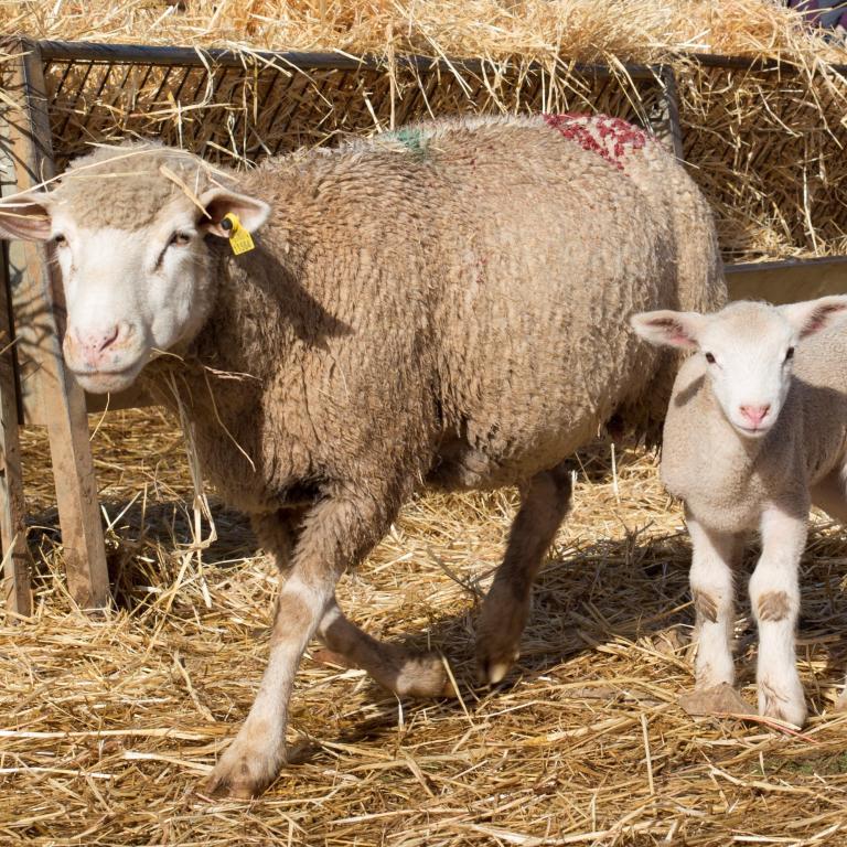
Får og geder
AETIOLOGY:
Mycoplasma agalactiae, Mycoplasma capricolum, Mycoplasma mycoides, Mycoplasma putrefaciens.
TRANSMISSION:
The main transmission method is via the ingestion of contaminated food and water and, sometimes, via the conjunctiva. The excretion routes are faeces, urine, nasal and conjunctival secretion, milk, abortions and open joints. Occasionally the disease is transmitted indirectly via fomites. Once the disease is present within a farm, it can stay there for a long time as an endemic disease. Generally low mortality.
Enters the body of the animal through ingestion, reaches and crosses the intestine, and reaches the blood; this results in early sepsis. At 24 h it is present in the spleen, pancreas, liver and brain. At 5 days the animals present with mastitis and infection of the joints (lameness), eye (conjunctivitis and keratitis) and the pregnant uterus (causing abortion).
CLINICAL SIGNS:
Contagious agalactia is known for its three characteristic signs:
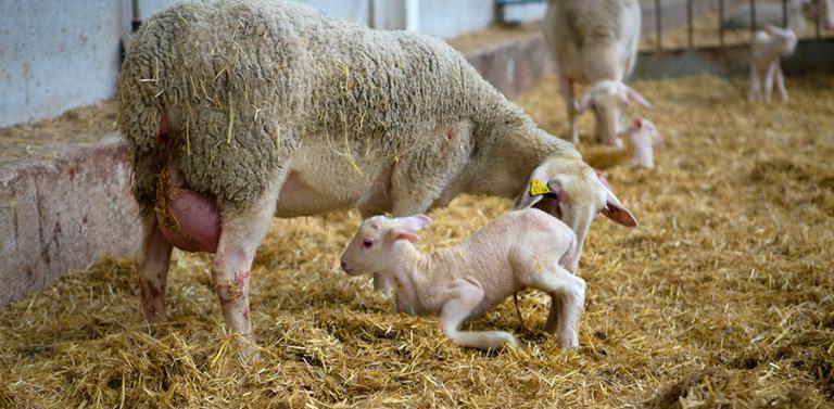
- Breast symptoms: the Mycoplasma destroys the glandular epithelium, causing agalactia. Mastitis that alters the appearance of milk (yellow, at rest in a green liquid fraction and a sediment).
- Joint symptoms: cartilage is eroded, and synovial fluid increases, causing arthritis. Lameness may occur due to inflammation or joints may ooze spontaneously and present fistulas, secreting a fibrinous exudate.
- Ocular symptoms: purulent and punctate inflammation in the form of conjunctivitis or keratitis (inflammation of the cornea) that impedes vision, leading to blindness.
- Other: occasional abortion when mycoplasmaemia coincides with the end of gestation, although the reproductive system is not targeted specifically. Depression, anorexia, hyperthermia of 41-42°C.
LESIONS:
- Udder: swelling and increase in size, grey and fragile parenchyma. Turbid milk.
- Eyes: whitish cornea due to the oedema, hyperaemia in the corneoscleral junction.
- Joints: thickening of the joint capsule and turbid synovial fluid.
DIAGNSOSIS:
- Clinical picture: presence of one or more types of these symptoms.
- Identification of the causative agent: isolation in milk samples, synovial fluid, abortion, etc.
- Serology: complement fixation test, ELISA (only in unvaccinated animals).
TREATMENT, PREVENTION AND CONTROL:
To control the disease it is necessary to vaccinate all animals with commercial inactivated vaccines, at least every six months. In order to attempt to eradicate the problem, isolate the infected animals and separate the offspring from the mother after birth, since infection occurs in the first days of life. An alternative is to keep two herds, one of which will be uninfected and the other one a carrier, making sure that very good biosecurity measures are in place first.
Relaterede produkter

Inaktiveret vaccine mod Chlamydia abortus og Salmonella Abortusovis hos får, i suspension til injektion.

Inaktiveret vaccine mod mastitis hos geder og får, som emulsion til injektion

Inaktiveret vaccine mod ondartet klovsyge hos får, som suspension til injektion

Inaktiveret vaccine mod enterotoksæmi, stivkrampe og miltbrandsemfysem hos drøvtryggere, som suspension til injektion

Inaktiveret vaccine mod enterotoksæmi, pludselig død, miltbrandsemfysem og nekrotis hepatitis hos drøvtyggere, som suspension til injektion

Inaktiveret vaccine mod smitsom agalakti, som suspension til injektion

Inaktiveret vaccine mod pasteurellose hos drøvtyggere, som suspension til injektion
Relaterede tjenester
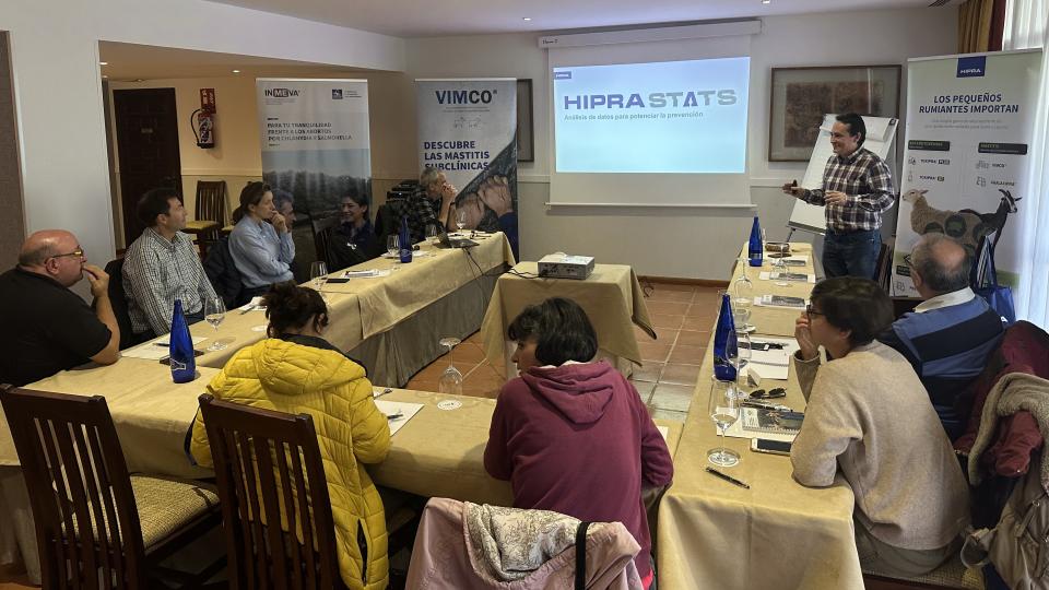
An innovative service based on data analysis that facilitates decision-making in small ruminants.

HIPRA Stats er en dataanalysetjeneste for dyreproduktionsvirksomheder.

HIPRA University tilbyder uddannelsesprogrammer af høj kvalitet inden for strategiske interesseområder til fagfolk

HIPRA har udviklet adskillige online-redskaber specielt til støtte for dyrlæger og andre fagfolk inden for husdyrproduktionen

Få adgang til en avanceret diagnostisk service med maksimal brugerkomfort og pålidelighed