Gå til hovedindhold
post mortem technique
Control of BVD and its impact in the herd: are we on the right path?
 

Disease dynamics 

BVDV cause both transient and persistent infections. The majority of transient infections of calves and non-pregnant adult cattle are subclinical, inducing immunosupression. Transient infection of naïve dams during pregnancy results in several severe effects depending on stages of gestation. If the dam becomes infected approximately between 30 and 125 days of gestation the calf will be born persistently infected. Other effects on reproductive performances are i.e. reduced conception rates, early embryonic death, abortion, birth of calves with congenital infection or physical malformations. Persistently infected (PI) animals are lifelong carriers and shedders of BVDV from all secretions, e.g., saliva, tears, milk, semen and faeces. PI animals do not produce antibodies against BVDV or produce a small amount due to their ability to seroconvert to strains of BVDV antigenically different to the persisting virus.  PI animals, if not removed from the herd, serve as a continuous source of the virus that perpetuate the disease in the herd and seroprevalence can reach 100% in less than two months for immunocompetent animals in direct contact with PIs. When infected animals come into contact with pregnant cattle, new PI calves may arise. Case-morbidity and fatality rates are very high in PI animals. Their lifespan is significantly shorter than other cattle in most cases, although not always, due to the increased likelihood of disease and poor performance or death (i.e. mucosal disease that presents as bloody diarrhea and ulcerated lesions). Case-morbidity and fatality rates may be increased in transient and persistent infections due to BVDV-induced immunosuppression and the increased susceptibility of infected animals to secondary infections, such as bovine respiratory disease complex (BRD) and enteric infections.  

Predisposing factor for BRD 

BRD is caused by a complex of viral and bacterial infection, linked to stressors related to management and environmental conditions. It is one of the most frequent disease that can affect young stock cattle including dairy, veal, preweaned beef, and feedlot calves. Weaning, crowding, sorting, commingling, processing, and shipping often trigger BRD.   

Viral involvement is considered as antecedent to, or concurrent with, bacterial infection. BVDV infection is a predisposing factor for BRD due to its known immunosuppressive effects on cells of both the innate and adaptive immune systems. Natural BRD outbreaks of feedlot cattle have highligheted the synergism between BVDV and bacterial infections. Several studies identified BVDV in affected calves and found that exposure to PI animals increased morbidity and risk of treatment for BRD (reviewed by Taylor et al., 2010). Other viruses are also implicated in BRD, i.e. bovine respiratory syncitial virus, bovine herpes virus 1, bovine parainfluenza type 3 virus and bovine respiratory coronavirus.  

Proper management of calves is of pivotal importance to minimize the risk of BRD and other pathogens and to improve their lifetime health and productivity. An adequate colostrum intake by calves, the hygiene and comfort of environment, proper cleaning and disinfection of calf feeding equipment, the use of calves-dedicated protective clothing and footwear prevent or decrease the spread of BVDV and other pathogens. On weaning and grouping in calf pens, the risk of exposure increases. Limiting the number of calves per pen, i.e. 7-10 animals, and segregation by similar age groups contribute to reduce the spread of pathogens, such as BVDV (Cannal et al., 2002). Moreover, it is important to avoid the commingling of weak and underweight animals that remain in pen as new groups arrive. Management and grouping size decisions must be made based on a balance between the risk and cost of disease versus the availability of facilities. 

BVD Technique postmortem

Clinical and laboratory diagnosis 

At present, BRD is most often identified based on clinical signs by human direct observation and through the use of imaging tecniques (Smith et al., 2020). However, monitoring changes in water or feed intake, animal movement and body temperature could allow the early detection of sub-clinical cattle. Taking into account the worldwide priority to reduce and rationalize antimicrobial use, an accurate clinical and laboratory diagnosis is increasingly required to identify pathogens involved in BRD and to optimally prevent and treat. The return on investment of laboratory diagnosis depends on selection of appropriate animals to sample, the sampling site of the respiratory tract and on the technical sampling skills of the veterinarian (Pardon & Buczinski, 2020). Deep nasopharyngeal swabs of animals in the first days of the disease, not previously treated with antimicrobials and not displaying severe respiratory signs, represent the first choice, taking into account age and pen grouping.  

Postmortem examination of lung reveals the distribution and texture of lesions, thereby suggesting causes of disease and providing tissues for confirmatory testing (Caswell et al., 2012). The multiple tissues must be sampled from the border between abnormal and normal lung and they should focus on primary lesions but also represent the spectrum of changes observed. The diagnostic investigation is invalidateded if the animals are sampled too late in the disease course and if specimens become autolyzed or undergo freeze-thaw damage. Postmortem examination must not only focus on the respiratory system but also search for clues in other tissues and organs. In particular for BVDV, oral and esophageal erosions and Peyer patch necrosis should be sought and additional samples, i.e. intestinal lesions and spleen, can be collected. Some PI animals may appear small, weak and ill-thrifty, showing a stunted growth and chronic ill thrift, therefore blood samples of these suspected PI animals have to be included. Among available diagnostic tests, multiplex polymerase chain reaction (PCR) for the diagnosis of BRD allows detection of multiple bacteria and viruses, providing practitioners test results within a day and allowing to target therapy or initiate control and prevention measures. Serologic tests are useful to target vaccination programs, to determine protective status, and to evaluate infection dynamics at larger scale (Pardon & Buczinski, 2020). 

Control and eradication approach 

Taking into account that BVDV control, unlike other infectious diseases involved in BRD, is highly feasible in cattle production systems and there are effective tools to break the disease transmission cycle, it is strongly recommended to establish the BVD status of the herd: whether there is evidence of BVD infection.  

In dairy herds, screening procedures to establish evidence of active BVD infection are based on the use of serology on a representative number of young animals (9 to 12 or 18 months of age); this approach gives an indication of ongoing or recent infection. If the BVD antibody level is negative in all the samples, it is very likely that they have not been exposed to BVDV recently. PCR can also be done on bulk tank milk for detection of viral circulation within the milking herd. If bulk milk tests positively, BVDV is circulating among the milking cows, but a negative test result is not proof of a BVDV free status.  

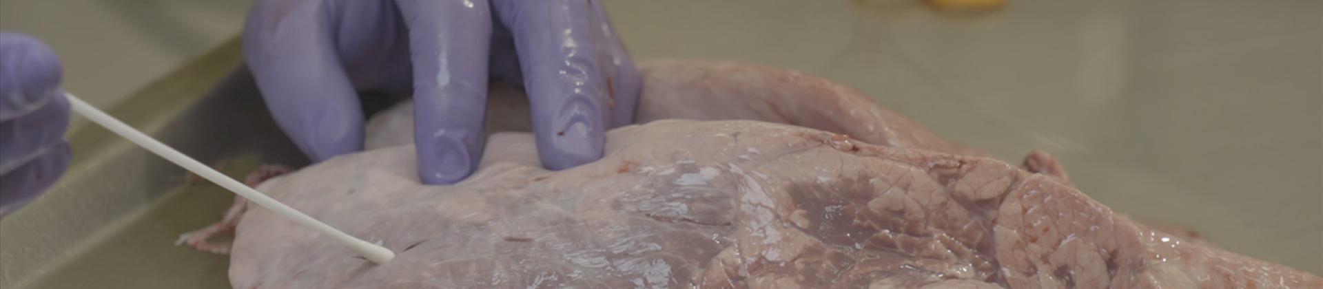
A second step is identification of individual PI animals on BVDV-positive farms. Testing of pooled blood samples by PCR is a rational means of excluding groups of animals from being BVDV carriers. The latter approach can be performed in combination with PCR on bulk milk as screening procedure in BVDV vaccinated herds. It is advisable to perform sampling and testing for PI animals also in newborns to early identify sources of BVDV infection. Due to colostral antibodies interference with detection of BVDV that might yield false-negative results in calves younger than 4-6 months of age, individual blood samples by PCR or skin biopsies (ear-notch samples) by ELISA may provide reliable test results (Sandvik et al., 2006). BVDV positive animals have to be retested after at least 3 weeks later, in order to rule out transient infection (Schweizer et al., 2021). 

The identification and removal of PI animals is considered key to control BVD and have a strongly beneficial impact on the welfare of cattle by preventing transient infections.  Central elements of systematic approaches to control and eradication of BVDV have been identified (Lindberg et al., 2006; EFSA AHAW Panel, 2017):  

  • Biosecurity involves all measures aimed at reducing between-herd transmission, with strong emphasis on preventing contacts with/movements of PI animals, i.e livestock trade, exhibitions and animal contacts on pasture. The efficacy of movement controls is most effective in the context of systematic control at regional and national level. The role of vaccines in systematic control is an additional biosecurity measure; vaccination aims to protect susceptible animals within herd after removal of PI animals or to prevent re-introduction of the infection in free herds, especially in areas where the risk of introducing BVDV infection is known or perceived to be high.   
  • The second measure is virus elimination by removal of PI animals from infected herds.   
  • Surveillance is fundamental to monitor the progress of interventions and to rapidly detect new infections.   


Author: Camilla Luzzago, DVM, PhD. Associate Professor of Animal Infectious Diseases. Department of Veterinary Medicine and Animal Science. Università degli Studi di Milano, Milan, Italy.


References: 

1. Bauermann, F.V., Flores, E.F., Ridpath, J.F. (2012) Antigenic relationships between Bovine viral diarrhea virus 1 and 2 and HoBi virus: possible impacts on diagnosis and control. J Vet Diagn Invest 24:253–261. doi.org/10.1177/1040638711435144 

2. Callan, R. J., & Garry, F. B. (2002). Biosecurity and bovine respiratory disease. The Veterinary Clinics of North America. Food Animal Practice, 18(1), 57–77. https://doi.org/10.1016/S0749-0720(02)00004-X 

3. Caswell, J.L., Hewson, J.   Slavić, D., DeLay, J., Bateman, K.,  (2012) Laboratory and Postmortem Diagnosis of Bovine Respiratory Disease Vet Clin North Am Food Anim Pract.  28(3): 419–441. doi: 10.1016/j.cvfa.2012.07.004 

4. EFSA AHAW Panel (EFSA Panel on Animal Health and Welfare), More S, Bøtner A, Butterworth A, Calistri P, Depner K, Edwards S, Garin-Bastuji B, Good M, Gort_azar Schmidt C, Michel V, Miranda MA, Nielsen SS, Raj M, Sihvonen L, Spoolder H, Stegeman JA, Thulke H-H, Velarde A, Willeberg P, Winckler C, Baldinelli F, Broglia A, Dhollander S, Beltr_an-Beck B, Kohnle L  and Bicout D (2017) Scientific Opinion on the assessment of listing and categorisation of animal diseases within the framework of the Animal Health Law (Regulation (EU) No 2016/429): bovine viral diarrhoea (BVD). EFSA Journal 2017;15(8):4952, 45 pp. https://doi.org/10.2903/j.efsa.2017.4952 

5. Lindberg A, Brownlie J, Gunn GJ, Houe H, Moennig V, Saatkamp HW, Sandvik T and Valle PS, (2006). The control of bovine viral diarrhoea virus in Europe: today and in the future. Revue Scientifique et Technique (International Office of Epizootics), 25, 961–979.  

6. Loneragan, G. H., Thomson, D. U., Montgomery, D. L., Mason, G. L., & Larson, R. L. (2005). Prevalence, outcome, and health consequences associated with persistent infection with bovine viral diarrhea virus in feedlot cattle. Journal of the American Veterinary Medical Association, 226(4), 595–601. https://doi.org/10.2460/javma.2005.226.595 

Moennig, V.; Yarnall, M.J. (2021) The Long Journey to BVD Eradication. Pathogens, 10, 1292. https://doi.org/10.3390/pathogens10101292 

Pardon, B. & Buczinski, S. (2020) Bovine Respiratory Disease Diagnosis What Progress Has Been Made in Infectious Diagnosis? Vet Clin Food Anim 36, 425–444 https://doi.org/10.1016/j.cvfa.2020.03.005 

Sandvik, T., Greiser-Wilke, I., Graham, D. Genome, diagnosis & diagnostic tools (2006) EU Thematic network on BVDV control position paper 

Schweizer, M., Stalder, H., Haslebacher, A., Grisiger, M., Schwermer, H., & Di Labio, E. (2021). Eradication of Bovine Viral Diarrhoea (BVD) in Cattle in Switzerland: Lessons Taught by the Complex Biology of the Virus. Frontiers in Veterinary Science, 8, 1012. https://doi.org/10.3389/fvets.2021.702730 

Smith, R. A., Step, D.L., Woolums, A. (2020) Bovine Respiratory Disease Looking Back and Looking Forward, What Do We See? Vet Clin Food Anim 36, 239–251 https://doi.org/10.1016/j.cvfa.2020.03.009 

Taylor, J. D., Fulton, R. W., Lehenbauer, T. W., Step, D. L., & Confer, A. W. (2010). The epidemiology of bovine respiratory disease: What is the evidence for predisposing factors? The Canadian Veterinary Journal, 51(10), 1095–1102. 

Yeşilbağ, K., Alpay, G., Becher, P. (2017) Variability and Global Distribution of Subgenotypes of Bovine Viral Diarrhea Virus. Viruses 9:128. doi.org/10.3390/v9060128