2a. Clostridium novyi Review (part 1)
กระทู้
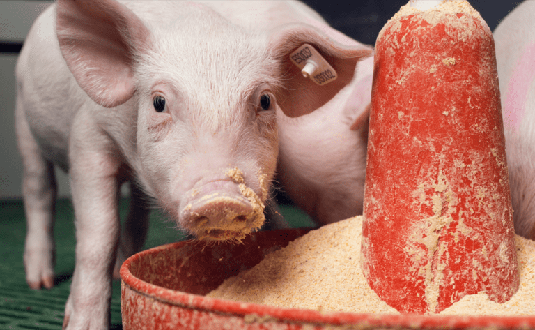
สุกร
Understanding what is Clostridium novyi
Clostridium novyi: Subtypes and toxins
Clostridia are Gram-positive, sporulating, oxygen-tolerant-to-strictly anaerobic rods, many of which cause disease in pigs, other domestic animals, and humans (Figure 1 and 2).
They can come from exogenous (most commonly the soil or animal feces) or endogenous sources (often the intestinal tract of the affected individual) (Songer, 1996).


Figure 1. Smears from the liver of a sow that died of Clostridium novyi infection, showing large gram-positive rods with oval to cylindrical subterminal spores (magnification ×1000). Source: Dr. Alfredo García Sánchez, Centro de Investigaciones Científicas y Tecnológicas de Extremadura (CICYTEX).


Figure 2. Microscopic lesions with Clostridium novyi infection. Gram and fluorescent antibody stains of direct smears and bacteriologic culture. Source: Dr. Roy Schultz, Iowa State University’s College of Veterinary Medicine.
Pathogenesis of most clostridial infections is based upon production of toxins, and can be categorized as neurotoxic, histotoxic, and enteric as seen in Table 1 (Hatheway, 1990).

Clostridium novyi was first isolated in the late 19th century (Novy, 1894). As noted (Table 2), it occurs as multiple types, differentiated by selective toxin production.
Differential production of alpha and beta toxins determines the toxin phenotype. Clostridium novyi type C is nontoxigenic (and therefore avirulent), but types A, B, and D (C. haemolyticum), cause disease in humans and domestic animals.
Thus, Clostridium novyi type A produces the organism’s alpha toxin. Type B, the primary cause of disease in sheep and swine, produces alpha toxin, but also makes a moderate amount of C. novyi beta toxin (Schranner et al, 1992). Finally, type D produces no alpha toxin, but secretes much more beta toxin than type B strains (Table 2).
It seems that C. novyi types B, C, and D are one independent species arising from the same phylogenetic background (Sasaki et al, 2002; Sasaki et al, 2001; Chen et al, 2011).

Table 2. C. novyi selective toxin production.
C. novyi in humans
Before discussing the specific impact of C. novyi on domestic animals and in particular on swine, it is probably worth emphasizing the role of C. novyi in human gas gangrene (Majumdar et al, 2004).
Dormant spores in muscles are potential sources of wound infections. Tyxpically, spores are introduced into a wound where they germinate and produce alpha toxin. Thus, the disease caused by C. novyi seems to be more common following wartime injuries than in those injuries experienced in peacetime.
There is no evidence that human infections are, strictly speaking, zoonotic, although the source of C. novyi may be, ultimately, the feces of domestic animals, which contaminate the environment.
C. novyi in other species
Disease in sheep and cattle
Formal infectious necrotic hepatitis in sheep is best known as an acute toxemia, in which disease begins in areas of liver necrosis caused by migration of liver flukes (Mitchell, 2002). Spore germination and organismal growth in these necrotic lesions leads to production and dissemination of alpha and beta toxins, and systemic toxin effects lead to the death of the animal.
Clostridium novyi infection in sheep and cattle is sometimes called “black disease”, due to accumulation of blood in the vasculature on the underside of the skin; it becomes black as clots develop and the blood lyses (Hamid et al, 1991; Duncan 1984).
Clostridium novyi type D causes bacillary hemoglobinuria of cattle and other ruminants. Type D spores found frequently in the intestinal tract cross the intestinal wall, enter portal circulation, and deposit in the liver. Immature flukes migrate through liver, causing necrosis and hypoxia and inducing germination of spores in Kupffer cells.
Beta toxin causes hepatic necrosis, and dissemination through the bloodstream causes intravascular hemolysis and hemorrhage. Clinical signs include fever, pale mucus membranes, anorexia, and abdominal pain. The case fatality rate is 90-95%.
Disease in pigs
There is no good information on the colonization rate in pigs of any age (sows, boars, piglets, fattening pigs). Disease, however, seems always of highest incidence by far among sows, suggesting a specific pathophysiologic event that predisposes sows to C. novyi hepatitis, toxemia, and bacteremia.
The disease has not been satisfactorily reproduced by inoculation of pigs with spores of C. novyi, but there is sufficient information from diagnostic work to conclude that C. novyi causes porcine infectious necrotic hepatitis. Deaths due to this condition are almost all focused in the peripartum period near the end of the sow cycle.
In cases of unexpected mortality in gestating sows, veterinarians need to be aware of the most common causes of death, including C. novyi infection. In order to achieve a correct diagnosis, it is essential to perform a postmortem examination and collect samples as soon as possible after death.
The need to be aware of porcine infectious necrotic hepatitis
There is nothing so disconcerting to a farmer or veterinarian on a pig farm as the sudden death of breeding sows.
On the one hand, they are animals in which a large amount has been invested and on the other, a high mortality rate increases the renewal rate and therefore the percentage of primparae, with a consequent decrease in the general productivity of the farm (García, 2013).
Pathogenesis of C. novyi infections and toxin action
The local lesion, whether in humans or domestic animals, is a zone of necrosis. Alpha-toxigenic C. novyi strains cause disease that presents with an archipelago of signs and symptoms, including refractory hypotension, leukocytosis, and various effusions (Ryan et al, 2001).
All of the large clostridial cytotoxins consist of single peptide chains averaging about 250,000 MW. Its action upon injection is characterized by fluid loss into the interstitial space.
Specifically, clostridial toxins glucosylate GTPases that are involved in actin cytoskeletal function impairing proper cell:cell contact (Selzer et al, 1996; Popoff and Bouvet, 2009), and leading to loss of integrity of vascular endothelium (Pires et al, 2012).
Epidemiology and transmission
It may be safe to say that, in the vast majority of cases, it is a peripartum disease. Rather, it is more probable that some physiologic event in proximity to parturition in the body of the sow is the inciting factor for disease.
Piglets could, conceivably, be colonized in the farrowing crate or stall during the first few hours after farrowing. In fact, it seems likely that exposure to a shedding sow early in life or to growing pigs at any stage is the most likely route to colonization.
The average parity of sows experiencing infectious necrotic hepatitis is about 5-6 litters. Sows in good body condition are at greatest risk, and disease occurs most often in the summer. Most cases occur in lactating sows and losses can easily exceed 15–30% per year.
Colonization without excretion (or simple shedding) seems a completely unreasonable expectation, so a colonized animal should be capable of spreading the organism to other pigs.
Clinical signs
The course of the disease is generally peracute, with hardly any clinical signs, which makes identification of sick animals difficult. The animals generally appear to be in good physical condition and then die suddenly, with unusual and rapid postmortem decomposition being characteristic (García, 2013).
Pathological changes include gross distention of the carcass, purple discoloration of the skin, generalized edema and subcutaneous infiltration with bubbles and foul smelling bloody fluid in pericardial, pleural, and abdominal cavities (Figure 3, A and B).
All organs are typically softened, spongy, gas-filled, and severely necrotized.


Figure 3. Image of a dead sow. Source: HIPRA.
Livers are enlarged and dark, and the parenchyma is uniformly infiltrated with gas bubbles, giving a spongy, “aerochocolate” appearance (Figure 4, A and B). Cases are sometimes accompanied by gastric ulcers.


Figure 4. The liver uniformly infiltrated with gas bubbles, presenting a spongy appearance on the cut surface, probably the most distinguishing feature of sudden death in sows caused by Clostridium novyi. Source: Dr. Alfredo García Sánchez, Centro de Investigaciones Científicas y Tecnológicas de Extremadura (CICYTEX).
Field case
Evidence of an outdoor pig-breeding unit of the Tierpark Arche Warder e. V. (Germany), where 16 pigs of different ages and sexes died in October 2011.
Necropsy revealed tympany, liver emphysema, subcutaneous edema, hemopericardium, hemothorax, and intense gas bubble infiltrations in muscles. The stomachs were filled. Initial anaerobic bacteriological investigations gave negative results, but PCR analysis of tissues revealed the flagellin gene of C. novyi types A and B.
Thus, C. novyi infection was diagnosed as the cause of the pig mortality (Jandowsky et al, 2013).
REFERENCES
• Anonymous. 2007. Defining sow productivity: sow mortality kills profits. J Swine Hlth Product. 15:295-296.
• Borrmann E and Schulze F. 1999. Detection of Clostridium novyi type B alpha toxin by cell culture systems. FEMS Immunol Med Microbiol. 24:275–280.
• Borrmann E, Schulze F, Cussler K, Hamel I, and Diller R. 2006. Development of a cell culture assay for the quantitative determination of vaccination-induced antibodies in rabbit sera against Clostridium perfringens epsilon toxin and Clostridium novyi alpha toxin. Vet Microbiol. 114:41-50.
• Chen Y, Indurthi DC, Jones SW, and Papoutsakis ET. 2011. Small RNAs in the genus Clostridium. MBio. 2:e00340-10.
• Duncan LF. 1984. Liver biopsy and black disease in a sheep. Aust Vet J. 61:272-273.
• Eklund MW, Poysky FT, Meyers JA, and Pelroy GA. 1974. Interspecies conversion of Clostridium botulinum type C to Clostridium novyi type A by bacteriophage. Science. 186:456-458.
• García A, Ayuso D, Benítez JM, García WL, Martínez R, and Sánchez S. 2009. Clostridium novyi infection causing sow mortality in an Iberian pig herd raised in an outdoor rearing system in Spain. J Swine Health Produc. 17:264-269.
• García A. 2013. Sanidad del ganado porcino ibérico. Principales enfermedades infecciosas. Librería Técnica Figueroa-2 (eds) PUBLICEP-Producción Humanes de Madrid, Madrid; pp 127-130.
• Hamid ME, Mohamed G, Abu Samra MT, and Hamad AA. 1991. First report of infectious necrotic hepatitis (black disease) among Nubian goats in Sudan. Rev Elev Med Vet Pays Trop. 44:273-275.
• Hatheway CL. 1990. Toxigenic clostridia. Clin Microbiol Rev. 3:66-98.
• Jandowsky A, Bodenthin A, Seyboldt C, and Frölich K. 2013. Sudden death of outdoor housed pigs caused by Clostridium novyi. A case report. Tierarztl Prax Ausg G Grosstiere Nutztiere. 41:392-395.
• Majumdar S, Woodcock S, and Cheesbrough J. 2004. Severe sepsis following wound infection by an unusual organism—Clostridium novyi. Int J Clin Pract. 58:892-893.
• McKenzie E. 2012. Clostridial myositis. Large Animal Proceedings. North American Veterinary Conference, Orlando, Florida, USA, 14-18 January 2012.
• Mitchell G. 2002. Update on fasciolosis in cattle and sheep. In Practice, 24: 378-385.
• Novy F. 1894. Ein neuer anaërober Bacillus des malignen Oedems. Medical Microbiol Immunol. 17:209-232.
• Pires PS, Ecco R, de Araujo MR, Silva ROS, Salavarani FM, Heneine LGD, Assis RA, and Lobato FCF. 2012. Comparative analysis of lesions caused by histotoxic clostridia in experimentally induced myonecrosis. Semina: Ciencias Agrarias (Londrina). 33:2337-2345.
• Popoff MR and Bouvet P. 2009. Clostridial toxins. Future Microbiol. 4:1021-1064.
• Poxton IR. 2007. A PCR approach to determine the distribution of toxin genes in closely related Clostridium species: Clostridium botulinum type C and D neurotoxins and C2 toxin, and Clostridium novyi alpha toxin. J Med Microbiol. 56:196-201.
• Ryan JM, Paul J, Curtis S, and Patel NK. 2001. Clostridium novyi infection: a fatal association with injecting drug users. Emerg Med J. 18:138-139.
• Sasaki Y, Takikawa N, Kojima A, Norimatsu M, Suzuki S, and Tamura Y. 2001. Phylogenetic positions of Clostridium novyi and Clostridium haemolyticum based on 16S rDNA sequences. Int J Syst Evol Microbiol. 51: 901-904.
• Sasaki Y, Kojima A, Aoki H, Ogikubo Y, Takikawa N, and Tamura Y. 2002. Phylogenetic analysis and PCR detection of Clostridium chauvoei, Clostridium haemolyticum, Clostridium novyi types A and B, and Clostridium septicum based on the flagellin gene. Vet Microbiol. 86:257-267.
• Schranner I, Erhard MH, Kaltner H, Lapsch U. 1992. Isolation of immunogenic and lethal peptides of alpha-toxin from Clostridium novyi type B. Toxicon. 30:653-668.
• Schultz R; Dau D, Hoefling D, Duran O, Carson T, BetonL, Woodward C, Pollard K, Busker K, Kaster D, and SteidingerM. 2001. A sow mortality study- the real reasons sows die: identifyingcauses and implementing action. Proceedings of the AmericanAssociation of Swine Veterinarians Ames, Iowa. 387-395.
• Selzer J, Hofmann F, Rex G, Wilm M, Mann M, Just I, and Aktories K. 1996. Clostridium novyi alpha-toxin-catalyzed incorporation of GlcNAc into Rho subfamily proteins. J Biol Chem. 271:25173-25177.
• Songer JG. 1996. Clostridial enteric diseases of domestic animals. Clin Microbiol Rev. 9:216-234.
ผลิตภัณฑ์ที่เกี่ยวข้อง

วัคซีนป้องกัน Mycoplasma hyopneumoniae และ PCV2 ในวัคซีนเดียวในสารแขวนลอยสำหรับฉีดเข้าใต้ผิวหนังแบบปราศจากเข็มตัวแรก

วัคซีนเชื้อเป็นสำหรับป้องกันโรคกลุ่มอาการทางระบบสืบพันธุ์และระบบทางเดินหายใจ (พีอาร์อาร์เอส) ในรูปผงแห้ง

สารเวอโรทอกซิน (verotoxin 2e) ชนิดเชื้อตายผ่านการกลั่น ป้องกันโรคบวมน้ำในยาแขวนตะกอนแบบฉีด

วัคซีนเชื้อตายสำหรับป้องกันโรคโพรงจมูกอักเสบแบบรุนแรงและไม่รุนแรงในสุกร ในรูปสารแขวนตะกอน
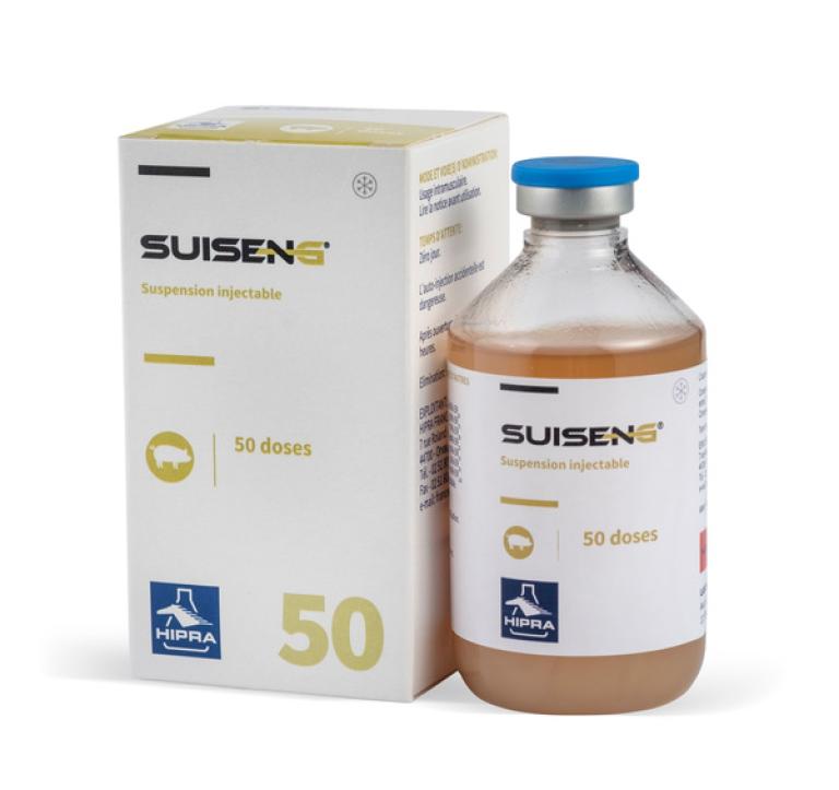
วัคซีนเชื้อตายสาหรับป้องกัน โรคโคไลบาซิลโลซิสในลูกสุกรแรกเกิด และโรคติดเชื้อ แบคทีเรีย คลอสติเดียมในสุกร ในรูปสารแขวนตะกอน

วัคซีนชนิดเชื้อตายป้องกันการติดเชื้อ Clostridioides difficile และ Clostridium perfringens ชนิดเอในสุกรในสารแขวนลอยสำหรับฉีด
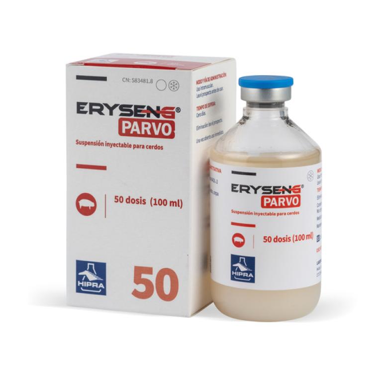
วัคซีนเชื้อตายสำหรับป้องกันโรคพาร์โวไวรัสในสุกร ในรูปสารแขวนตะกอน
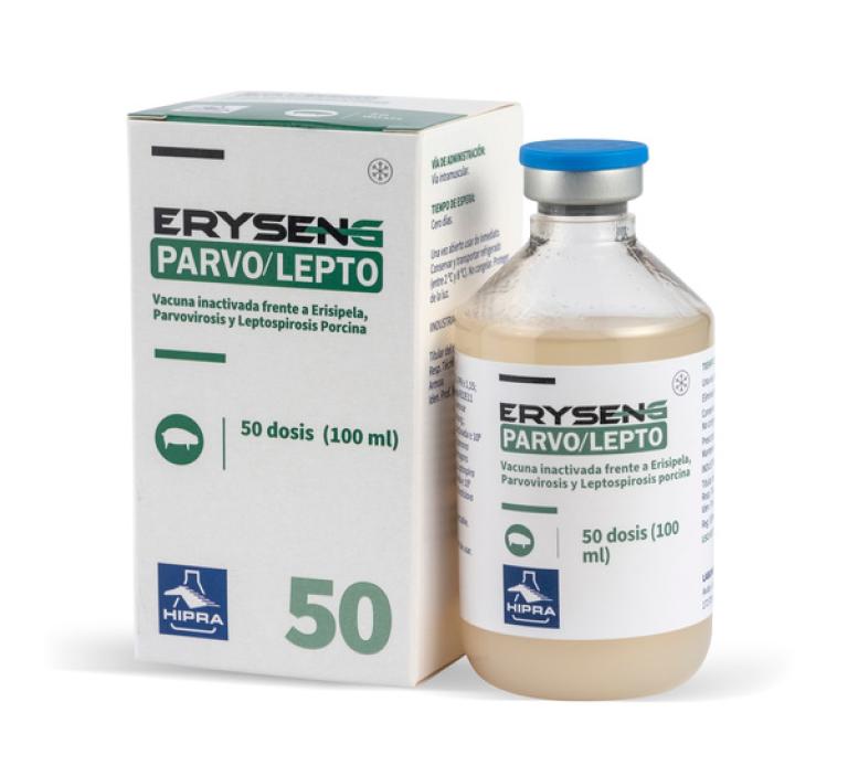
วัคซีนเชื้อตายสำหรับป้องกันโรคไข้หนังแดง โรคพาร์โวไวรัส และเลปโตสไปโรซิส ในรูปสารแขวนตะกอน

วัคซีนเชื้อเป็นสำหรับป้องกันโรคพิษสุนัขบ้าเทียม สายพันธุ์ที่มีการตัดยีนส่วน gE ในรูปผงแห้ง

วัคซีนเชื้อตายสำหรับป้องกันโรคไข้หนังแดงสุกร ในรูปสารแขวนตะกอน

วัคซีนเชื้อตายสำหรับป้องกันโรคพาร์โวไวรัสสุกร ในรูปสารแขวนตะกอน

วัคซีนเชื้อตายสาหรับป้องกัน โรคเอ็นซูติกนิวโมเนีย หรือโรคปอดอักเสบจากเชื้อมัยโคพลาสมา ในรูปสารแขวนตะกอน
บริการที่เกี่ยวข้อง

การทำวัคซีนอันชาญฉลาด (สมาร์ทวัคซิเนชัน)โดยฮิปรา เป็นแนวคิดวิวัฒนาการใหม่ที่รวมวิธีการทำวัคซีนอย่างชาญฉลาด เช่น เทคโนโลยีการติดฉลากเพื่อระบุสิ่งต่างๆโดยอาศัยคลื่นวิทยุ (RFID) เครื่องมือที่มีความแม่นยำและมีประสิทธิภาพสูงสำหรับการทำวัคซีน และการแก้ปัญหาเชิงดิจิตอลด้วยโปรแกรม ฮิปราลิงค์® ที่พัฒนาโดยบริษัทฮิปรา

การทำวัคซีนอันชาญฉลาด (สมาร์ทวัคซิเนชัน) สำหรับสุกรนำโดยนวัตกรรมวัคซีนยูนิสเตรน® พีอาร์อาร์เอส อุปกรณ์ฮิปราเดอมิค® และแอพพลิเคชันฮิปราลิงค์® ที่ทันสมัยสำหรับสัตวแพทย์มืออาชีพ
An innovative service based on data analysis that facilitates decision-making in swine

HIPRA Stats เป็นบริการวิเคราะห์ข้อมูลสําหรับบริษัทการผลิตสัตว์

Hipradermic® เป็นอุปกรณ์ฉีดยาขั้นสูงแบบไร้เข็ม พร้อมการเชื่อมต่อแบบไร้สายสําหรับการฉีดวัคซีนในผิวหนังของสุกร

ฮิปรา ยูนิเวอร์ซิตี้ มีโปรแกรมการฝึกอบรมคุณภาพสูงที่เป็นที่สนใจของมืออาชีพทุกท่าน

เข้าสู่ระบบการให้บริการด้านงานวินิจฉัยโรคสัตว์ด้วยความสะดวกและน่าเชื่อถือ